2VAL

Crystal structure of an Escherichia coli tRNAGly microhelix at 2.0 Angstrom resolution


Experimental Data Snapshot

  • Method: X-RAY DIFFRACTION
  • Resolution: 2.00 Å
  • R-Value Free: 0.318 
  • R-Value Work: 0.241 

wwPDB Validation   3D Report Full Report


This is version 1.4 of the entry. See complete history


Literature

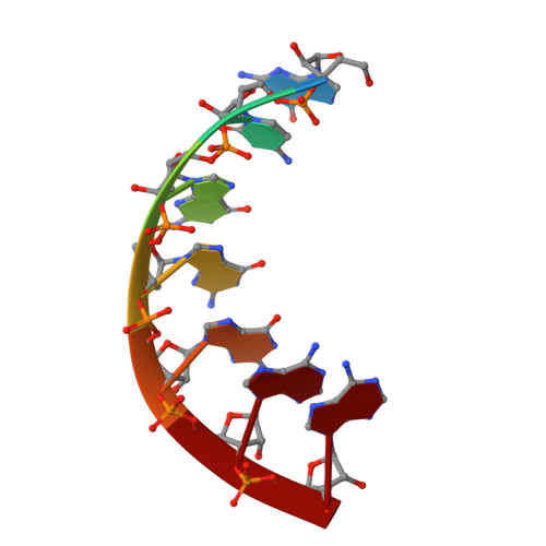
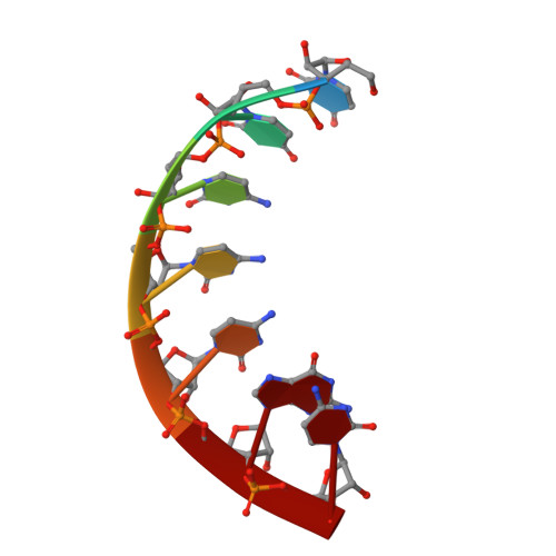
Crystal Structure of an Escherichia Coli Trnagly Microhelix at 2.0 Angstrom Resolution

Forster, C.Brauer, A.B.E.Perbandt, M.Lehmann, D.Furste, J.P.Betzel, C.Erdmann, V.A.

(2007) Biochem Biophys Res Commun 363: 621

  • DOI: https://doi.org/10.1016/j.bbrc.2007.09.008
  • Primary Citation of Related Structures:  
    2VAL

  • PubMed Abstract: 

    tRNA identity elements determine the correct aminoacylation by the cognate aminoacyl-tRNA synthetase. In class II aminoacyl tRNA synthetase systems, tRNA specificity is assured by rather few and simple recognition elements, mostly located in the acceptor stem of the tRNA. Here we present the crystal structure of an Escherichia coli tRNA(Gly) aminoacyl stem microhelix at 2.0 A resolution. The tRNA(Gly) microhelix crystallizes in the space group P3(2)21 with the cell constants a=b=35.35 A, c=130.82 A, gamma=120 degrees . The helical parameters, solvent molecules and a potential magnesium binding site are discussed.


  • Organizational Affiliation

    Institute of Chemistry and Biochemistry, Free University Berlin, Thielallee 63, 14195 Berlin, Germany.


Macromolecules

Find similar nucleic acids by:  Sequence   |   3D Structure  

Entity ID: 1
MoleculeChains LengthOrganismImage
5'-R(*GP*CP*GP*GP*GP*AP*AP)-3'
A, B
7Escherichia coli
Sequence Annotations
Expand
  • Reference Sequence

Find similar nucleic acids by:  Sequence   |   3D Structure  

Entity ID: 2
MoleculeChains LengthOrganismImage
5'-R(*UP*UP*CP*CP*CP*GP*CP)-3'
C, D
7Escherichia coli
Sequence Annotations
Expand
  • Reference Sequence
Small Molecules
Ligands 1 Unique
IDChains Name / Formula / InChI Key2D Diagram3D Interactions
MG
Query on MG

Download Ideal Coordinates CCD File 
E [auth C]MAGNESIUM ION
Mg
JLVVSXFLKOJNIY-UHFFFAOYSA-N
Experimental Data & Validation

Experimental Data

  • Method: X-RAY DIFFRACTION
  • Resolution: 2.00 Å
  • R-Value Free: 0.318 
  • R-Value Work: 0.241 
  • Space Group: P 32 2 1
Unit Cell:
Length ( Å )Angle ( ˚ )
a = 35.348α = 90
b = 35.348β = 90
c = 130.821γ = 120
Software Package:
Software NamePurpose
REFMACrefinement
PHASERphasing

Structure Validation

View Full Validation Report


Entry History 

Deposition Data

Revision History  (Full details and data files)

  • Version 1.0: 2007-10-16
    Type: Initial release
  • Version 1.1: 2011-05-08
    Changes: Version format compliance
  • Version 1.2: 2011-07-13
    Changes: Version format compliance
  • Version 1.3: 2019-07-24
    Changes: Data collection
  • Version 1.4: 2019-10-16
    Changes: Data collection, Other