2UYK

Crystal structure of E. coli TdcF with bound serine


Experimental Data Snapshot

  • Method: X-RAY DIFFRACTION
  • Resolution: 1.60 Å
  • R-Value Free: 0.199 
  • R-Value Work: 0.155 
  • R-Value Observed: 0.157 

wwPDB Validation   3D Report Full Report


This is version 1.3 of the entry. See complete history


Literature

The Crystal Structure of Escherichia Coli Tdcf, a Member of the Highly Conserved Yjgf/Yer057C/Uk114 Family.

Burman, J.D.Stevenson, C.E.M.Sawers, R.G.Lawson, D.M.

(2007) BMC Struct Biol 7: 30

  • DOI: https://doi.org/10.1186/1472-6807-7-30
  • Primary Citation of Related Structures:  
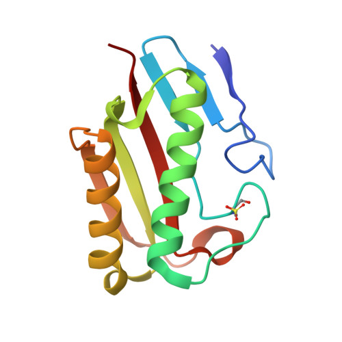
    2UYJ, 2UYK, 2UYN, 2UYP

  • PubMed Abstract: 

    The YjgF/YER057c/UK114 family of proteins is widespread in nature, but has as yet no clearly defined biological role. Members of the family exist as homotrimers and are characterised by intersubunit clefts that are delineated by well-conserved residues; these sites are likely to be of functional significance, yet catalytic activity has never been detected for any member of this family. The gene encoding the TdcF protein of E. coli, a YjgF/YER057c/UK114 family member, resides in an operon that strongly suggests a role in the metabolism of 2-ketobutyrate for this protein.


  • Organizational Affiliation

    Department of Biological Chemistry, John Innes Centre, Norwich, UK. j.burman@bath.ac.uk


Macromolecules
Find similar proteins by:  (by identity cutoff)  |  3D Structure
Entity ID: 1
MoleculeChains Sequence LengthOrganismDetailsImage
PROTEIN TDCF
A, B, C
129Escherichia coliMutation(s): 0 
UniProt
Find proteins for P0AGL2 (Escherichia coli (strain K12))
Explore P0AGL2 
Go to UniProtKB:  P0AGL2
Entity Groups  
Sequence Clusters30% Identity50% Identity70% Identity90% Identity95% Identity100% Identity
UniProt GroupP0AGL2
Sequence Annotations
Expand
  • Reference Sequence
Small Molecules
Ligands 1 Unique
IDChains Name / Formula / InChI Key2D Diagram3D Interactions
SER
Query on SER

Download Ideal Coordinates CCD File 
D [auth C]SERINE
C3 H7 N O3
MTCFGRXMJLQNBG-REOHCLBHSA-N
Modified Residues  1 Unique
IDChains TypeFormula2D DiagramParent
OCS
Query on OCS
A, B, C
L-PEPTIDE LINKINGC3 H7 N O5 SCYS
Experimental Data & Validation

Experimental Data

  • Method: X-RAY DIFFRACTION
  • Resolution: 1.60 Å
  • R-Value Free: 0.199 
  • R-Value Work: 0.155 
  • R-Value Observed: 0.157 
  • Space Group: P 21 21 2
Unit Cell:
Length ( Å )Angle ( ˚ )
a = 72.685α = 90
b = 86.351β = 90
c = 62.598γ = 90
Software Package:
Software NamePurpose
REFMACrefinement
DENZOdata reduction
SCALEPACKdata scaling
AMoREphasing

Structure Validation

View Full Validation Report


Entry History 

Deposition Data

Revision History  (Full details and data files)

  • Version 1.0: 2007-05-29
    Type: Initial release
  • Version 1.1: 2011-05-08
    Changes: Version format compliance
  • Version 1.2: 2011-07-13
    Changes: Version format compliance
  • Version 1.3: 2023-12-13
    Changes: Data collection, Database references, Derived calculations, Other, Refinement description