A buried lysine that titrates with a normal pKa: role of conformational flexibility at the protein-water interface as a determinant of pKa values.
Harms, M.J., Schlessman, J.L., Chimenti, M.S., Sue, G.R., Damjanovic, A., Garcia-Moreno, B.(2008) Protein Sci 17: 833-845
- PubMed: 18369193
- DOI: https://doi.org/10.1110/ps.073397708
- Primary Citation of Related Structures:
2RKS - PubMed Abstract:
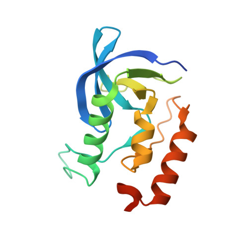
Previously we reported that Lys, Asp, and Glu residues at positions 66 and 92 in staphylococcal nuclease (SNase) titrate with pK(a) values shifted by up to 5 pK(a) units in the direction that promotes the neutral state. In contrast, the internal Lys-38 in SNase titrates with a normal pK(a). The crystal structure of the L38K variant shows that the side chain of Lys-38 is buried. The ionizable moiety is approximately 7 A from solvent and ion paired with Glu-122. This suggests that the pK(a) value of Lys-38 is normal because the energetic penalty for dehydration is offset by a favorable Coulomb interaction. However, the pK(a) of Lys-38 was also normal when Glu-122 was replaced with Gln or with Ala. Continuum electrostatics calculations were unable to reproduce the pK(a) of Lys-38 unless the protein was treated with an artificially high dielectric constant, consistent with structural reorganization being responsible for the normal pK(a) value of Lys-38. This reorganization must be local because circular dichroism and NMR spectroscopy indicate that the L38K protein is native-like under all conditions studied. In molecular dynamics simulations, the ion pair between Lys-38 and Glu-122 is unstable. The simulations show that a minor rearrangement of a loop is sufficient to allow penetration of water to the amino moiety of Lys-38. This illustrates both the important roles of local flexibility and water penetration as determinants of pK(a) values of ionizable groups buried near the protein-water interface, and the challenges faced by structure-based pK(a) calculations in reproducing these effects.
Organizational Affiliation:
Department of Biophysics, Johns Hopkins University, Baltimore, Maryland 21218, USA.