2REN

STRUCTURE OF RECOMBINANT HUMAN RENIN, A TARGET FOR CARDIOVASCULAR-ACTIVE DRUGS, AT 2.5 ANGSTROMS RESOLUTION


Experimental Data Snapshot

  • Method: X-RAY DIFFRACTION
  • Resolution: 2.50 Å
  • R-Value Observed: 0.217 

wwPDB Validation   3D Report Full Report


This is version 1.4 of the entry. See complete history


Literature

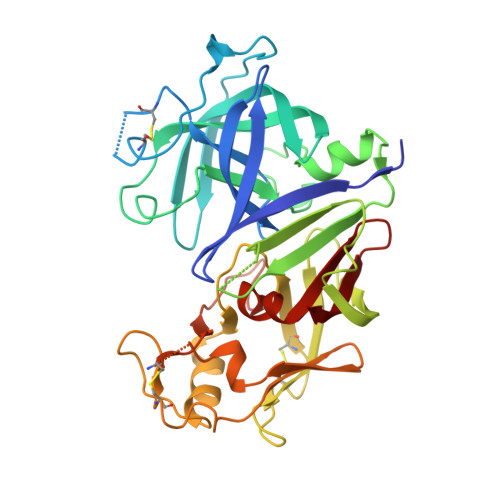
Structure of recombinant human renin, a target for cardiovascular-active drugs, at 2.5 A resolution.

Sielecki, A.R.Hayakawa, K.Fujinaga, M.Murphy, M.E.Fraser, M.Muir, A.K.Carilli, C.T.Lewicki, J.A.Baxter, J.D.James, M.N.

(1989) Science 243: 1346-1351

  • DOI: https://doi.org/10.1126/science.2493678
  • Primary Citation of Related Structures:  
    2REN

  • PubMed Abstract: 

    The x-ray crystal structure of recombinant human renin has been determined. Molecular dynamics techniques that included crystallographic data as a restraint were used to improve an initial model based on porcine pepsinogen. The present agreement factor for data from 8.0 to 2.5 angstroms (A) is 0.236. Some of the surface loops are poorly determined, and these disordered regions border a 30 A wide solvent channel. Comparison of renin with other aspartyl proteinases shows that, although the structural cores and active sites are highly conserved, surface residues, some of which are critical for specificity, vary greatly (up to 10A). Knowledge of the actual structure, as opposed to the use of models based on related enzymes, should facilitate the design of renin inhibitors.


  • Organizational Affiliation

    Department of Biochemistry, University of Alberta, Edmonton, Canada.


Macromolecules
Find similar proteins by:  (by identity cutoff)  |  3D Structure
Entity ID: 1
MoleculeChains Sequence LengthOrganismDetailsImage
RENIN340Homo sapiensMutation(s): 0 
EC: 3.4.23.15
UniProt & NIH Common Fund Data Resources
Find proteins for P00797 (Homo sapiens)
Explore P00797 
Go to UniProtKB:  P00797
PHAROS:  P00797
GTEx:  ENSG00000143839 
Entity Groups  
Sequence Clusters30% Identity50% Identity70% Identity90% Identity95% Identity100% Identity
UniProt GroupP00797
Sequence Annotations
Expand
  • Reference Sequence
Small Molecules
Ligands 1 Unique
IDChains Name / Formula / InChI Key2D Diagram3D Interactions
NAG
Query on NAG

Download Ideal Coordinates CCD File 
B [auth A]2-acetamido-2-deoxy-beta-D-glucopyranose
C8 H15 N O6
OVRNDRQMDRJTHS-FMDGEEDCSA-N
Experimental Data & Validation

Experimental Data

  • Method: X-RAY DIFFRACTION
  • Resolution: 2.50 Å
  • R-Value Observed: 0.217 
  • Space Group: I 4
Unit Cell:
Length ( Å )Angle ( ˚ )
a = 134.08α = 90
b = 134.08β = 90
c = 41.98γ = 90
Software Package:
Software NamePurpose
X-PLORmodel building
GROMOSrefinement
PROLSQrefinement
X-PLORrefinement
X-PLORphasing

Structure Validation

View Full Validation Report


Entry History 

Deposition Data

Revision History  (Full details and data files)

  • Version 1.0: 1994-01-31
    Type: Initial release
  • Version 1.1: 2008-03-25
    Changes: Version format compliance
  • Version 1.2: 2011-07-13
    Changes: Non-polymer description, Version format compliance
  • Version 1.3: 2017-11-29
    Changes: Derived calculations, Other
  • Version 1.4: 2020-07-29
    Type: Remediation
    Reason: Carbohydrate remediation
    Changes: Advisory, Data collection, Derived calculations, Structure summary