2R9Z

Glutathione amide reductase from Chromatium gracile


Experimental Data Snapshot

  • Method: X-RAY DIFFRACTION
  • Resolution: 2.10 Å
  • R-Value Free: 0.246 
  • R-Value Work: 0.197 
  • R-Value Observed: 0.199 

wwPDB Validation   3D Report Full Report


Ligand Structure Quality Assessment 


This is version 1.2 of the entry. See complete history


Literature

Understanding nicotinamide dinucleotide cofactor and substrate specificity in class I flavoprotein disulfide oxidoreductases: crystallographic analysis of a glutathione amide reductase.

Van Petegem, F.De Vos, D.Savvides, S.Vergauwen, B.Van Beeumen, J.

(2007) J Mol Biol 374: 883-889

  • DOI: https://doi.org/10.1016/j.jmb.2007.09.072
  • Primary Citation of Related Structures:  
    2R9Z, 2RAB

  • PubMed Abstract: 

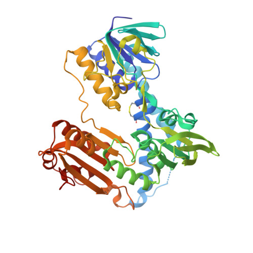
    Glutathione reductase (GR) plays a vital role in maintaining the antioxidant levels of the cytoplasm by catalyzing the reduction of glutathione disulfide to reduced glutathione, thereby using NADPH and flavin adenine dinucleotide as cofactors. Chromatiaceae have evolved an unusual homolog that prefers both a modified substrate (glutathione amide disulfide [GASSAG]) and a different cofactor (NADH). Herein, we present the crystal structure of the Chromatium gracile glutathione amide reductase (GAR) both alone and in complex with NAD(+). An altered charge distribution in the GASSAG binding pocket explains the difference in substrate specificity. The NADH binding pocket of GAR differs from that of wild-type GR as well as that of a low active GR that was engineered to mimic NADH binding. Based on the GAR structure, we propose two attractive rationales for producing an efficient GR enzyme with NADH specificity.


  • Organizational Affiliation

    Laboratory of Protein Biochemistry and Protein Engineering, Ghent University, 9000 Ghent, Belgium. filip.vanpetegem@gmail.com


Macromolecules
Find similar proteins by:  (by identity cutoff)  |  3D Structure
Entity ID: 1
MoleculeChains Sequence LengthOrganismDetailsImage
Glutathione amide reductase
A, B
463Marichromatium gracileMutation(s): 0 
UniProt
Find proteins for D0VWY5 (Marichromatium gracile)
Explore D0VWY5 
Go to UniProtKB:  D0VWY5
Entity Groups  
Sequence Clusters30% Identity50% Identity70% Identity90% Identity95% Identity100% Identity
UniProt GroupD0VWY5
Sequence Annotations
Expand
  • Reference Sequence
Small Molecules
Ligands 3 Unique
IDChains Name / Formula / InChI Key2D Diagram3D Interactions
FAD
Query on FAD

Download Ideal Coordinates CCD File 
C [auth A],
J [auth B]
FLAVIN-ADENINE DINUCLEOTIDE
C27 H33 N9 O15 P2
VWWQXMAJTJZDQX-UYBVJOGSSA-N
NI
Query on NI

Download Ideal Coordinates CCD File 
F [auth A],
H [auth A],
M [auth B],
O [auth B]
NICKEL (II) ION
Ni
VEQPNABPJHWNSG-UHFFFAOYSA-N
CL
Query on CL

Download Ideal Coordinates CCD File 
D [auth A]
E [auth A]
G [auth A]
I [auth A]
K [auth B]
D [auth A],
E [auth A],
G [auth A],
I [auth A],
K [auth B],
L [auth B],
N [auth B],
P [auth B]
CHLORIDE ION
Cl
VEXZGXHMUGYJMC-UHFFFAOYSA-M
Experimental Data & Validation

Experimental Data

  • Method: X-RAY DIFFRACTION
  • Resolution: 2.10 Å
  • R-Value Free: 0.246 
  • R-Value Work: 0.197 
  • R-Value Observed: 0.199 
  • Space Group: P 41
Unit Cell:
Length ( Å )Angle ( ˚ )
a = 71.933α = 90
b = 71.933β = 90
c = 223.854γ = 90
Software Package:
Software NamePurpose
REFMACrefinement
DENZOdata reduction
SCALEPACKdata scaling
AMoREphasing

Structure Validation

View Full Validation Report


Ligand Structure Quality Assessment 


Entry History 

Deposition Data

Revision History  (Full details and data files)

  • Version 1.0: 2008-02-19
    Type: Initial release
  • Version 1.1: 2011-07-13
    Changes: Version format compliance
  • Version 1.2: 2023-08-30
    Changes: Data collection, Database references, Derived calculations, Refinement description