NMR structure of the protein NP_247299.1: comparison with the crystal structure.
Jaudzems, K., Geralt, M., Serrano, P., Mohanty, B., Horst, R., Pedrini, B., Elsliger, M.A., Wilson, I.A., Wuthrich, K.(2010) Acta Crystallogr Sect F Struct Biol Cryst Commun 66: 1367-1380
- PubMed: 20944234
- DOI: https://doi.org/10.1107/S1744309110005890
- Primary Citation of Related Structures:
2KLA, 2QTD - PubMed Abstract:
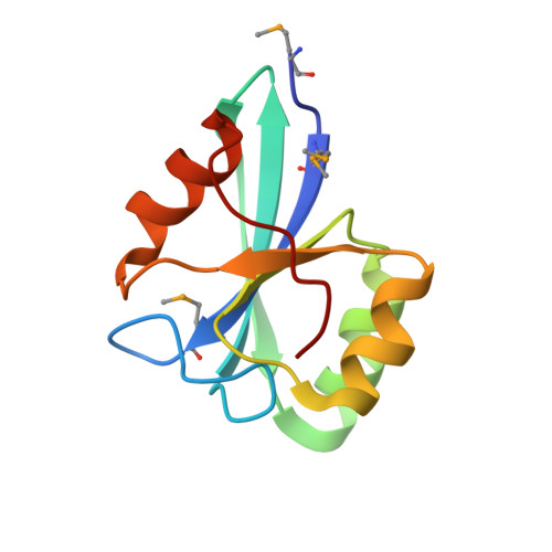
The NMR structure of the protein NP_247299.1 in solution at 313 K has been determined and is compared with the X-ray crystal structure, which was also solved in the Joint Center for Structural Genomics (JCSG) at 100 K and at 1.7 Å resolution. Both structures were obtained using the current largely automated crystallographic and solution NMR methods used by the JCSG. This paper assesses the accuracy and precision of the results from these recently established automated approaches, aiming for quantitative statements about the location of structure variations that may arise from either one of the methods used or from the different environments in solution and in the crystal. To evaluate the possible impact of the different software used for the crystallographic and the NMR structure determinations and analysis, the concept is introduced of reference structures, which are computed using the NMR software with input of upper-limit distance constraints derived from the molecular models representing the results of the two structure determinations. The use of this new approach is explored to quantify global differences that arise from the different methods of structure determination and analysis versus those that represent interesting local variations or dynamics. The near-identity of the protein core in the NMR and crystal structures thus provided a basis for the identification of complementary information from the two different methods. It was thus observed that locally increased crystallographic B values correlate with dynamic structural polymorphisms in solution, including that the solution state of the protein involves a slow dynamic equilibrium on a time scale of milliseconds or slower between two ensembles of rapidly interchanging conformers that contain, respectively, the cis or trans form of the C-terminal proline and represent about 25 and 75% of the total protein.
Organizational Affiliation:
Department of Molecular Biology, The Scripps Research Institute, La Jolla, CA 92037, USA.