2QT7

Crystallographic structure of the mature ectodomain of the human receptor-type protein-tyrosine phosphatase IA-2 at 1.30 Angstroms


Experimental Data Snapshot

  • Method: X-RAY DIFFRACTION
  • Resolution: 1.30 Å
  • R-Value Free: 0.242 
  • R-Value Work: 0.220 
  • R-Value Observed: 0.222 

wwPDB Validation   3D Report Full Report


This is version 1.3 of the entry. See complete history


Literature

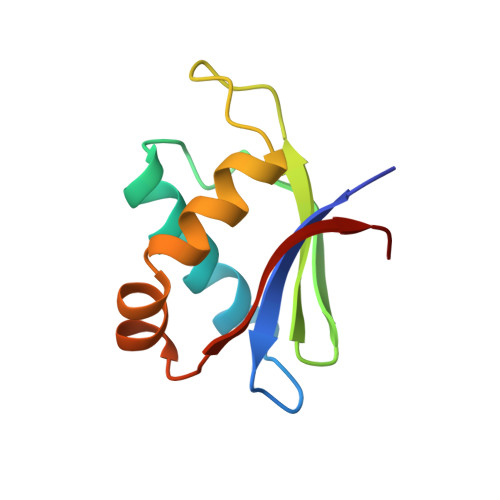
Structure of the Mature Ectodomain of the Human Receptor-type Protein-tyrosine Phosphatase IA-2

Primo, M.E.Klinke, S.Sica, M.P.Goldbaum, F.A.Jakoncic, J.Poskus, E.Ermacora, M.R.

(2008) J Biol Chem 283: 4674-4681

  • DOI: https://doi.org/10.1074/jbc.M708144200
  • Primary Citation of Related Structures:  
    2QT7, 3N4W, 3NG8

  • PubMed Abstract: 

    IA-2 (insulinoma-associated protein 2) is a protein-tyrosine phosphatase receptor located in secretory granules of neuroendocrine cells. Initially, it attracted attention due to its involvement in the autoimmune response associated to diabetes. Later it was found that upon exocytosis, the cytoplasmic domain of IA-2 is cleaved and relocated to the nucleus, where it enhances the transcription of the insulin gene. A concerted functioning of the whole receptor is to be expected. However, very little is known about the structure and function of the transmembrane and extracellular domains of IA-2. To address this issue, we solved the x-ray structure of the mature ectodomain of IA-2 (meIA-2) to 1.30A resolution. The fold of meIA-2 is related to the SEA (sea urchin sperm protein, enterokinase, agrin)) domains of mucins, suggesting its participation in adhesive contacts to the extracellular matrix and providing clues on how this kind of molecule may associate and form homo- and heterodimers. Moreover, we discovered that meIA-2 is self-proteolyzed in vitro by reactive oxygen species, suggesting the possibility of a new shedding mechanism that might be significant in normal function or pathological processes. Knowledge of meIA-2 structure should facilitate the search of its possible ligands and molecular interactions.


  • Organizational Affiliation

    Consejo Nacional de Investigaciones Científicas y Técnicas (Conicet), Rivadavia 1917 C1033AAJ, Ciudad Autónoma de Buenos Aires, Argentina.


Macromolecules
Find similar proteins by:  (by identity cutoff)  |  3D Structure
Entity ID: 1
MoleculeChains Sequence LengthOrganismDetailsImage
Receptor-type tyrosine-protein phosphatase-like N
A, B
91Homo sapiensMutation(s): 0 
Gene Names: PTPRNICA3ICA512
EC: 3.1.3.48
UniProt & NIH Common Fund Data Resources
Find proteins for Q16849 (Homo sapiens)
Explore Q16849 
Go to UniProtKB:  Q16849
PHAROS:  Q16849
GTEx:  ENSG00000054356 
Entity Groups  
Sequence Clusters30% Identity50% Identity70% Identity90% Identity95% Identity100% Identity
UniProt GroupQ16849
Sequence Annotations
Expand
  • Reference Sequence
Experimental Data & Validation

Experimental Data

  • Method: X-RAY DIFFRACTION
  • Resolution: 1.30 Å
  • R-Value Free: 0.242 
  • R-Value Work: 0.220 
  • R-Value Observed: 0.222 
  • Space Group: P 21 21 21
Unit Cell:
Length ( Å )Angle ( ˚ )
a = 31.756α = 90
b = 66.799β = 90
c = 73.131γ = 90
Software Package:
Software NamePurpose
REFMACrefinement
Blu-Icedata collection
HKL-2000data reduction
HKL-2000data scaling
SHELXDEphasing

Structure Validation

View Full Validation Report


Entry History 

Deposition Data

Revision History  (Full details and data files)

  • Version 1.0: 2007-12-11
    Type: Initial release
  • Version 1.1: 2011-07-13
    Changes: Version format compliance
  • Version 1.2: 2017-10-25
    Changes: Refinement description
  • Version 1.3: 2024-02-21
    Changes: Data collection, Database references, Derived calculations