2PV9

Crystal structure of murine thrombin in complex with the extracellular fragment of murine PAR4


Experimental Data Snapshot

  • Method: X-RAY DIFFRACTION
  • Resolution: 3.50 Å
  • R-Value Free: 0.319 
  • R-Value Work: 0.306 
  • R-Value Observed: 0.306 

wwPDB Validation   3D Report Full Report


Ligand Structure Quality Assessment 


This is version 1.5 of the entry. See complete history


Literature

Crystal structures of murine thrombin in complex with the extracellular fragments of murine protease-activated receptors PAR3 and PAR4.

Bah, A.Chen, Z.Bush-Pelc, L.A.Mathews, F.S.Di Cera, E.

(2007) Proc Natl Acad Sci U S A 104: 11603-11608

  • DOI: https://doi.org/10.1073/pnas.0704409104
  • Primary Citation of Related Structures:  
    2PUX, 2PV9

  • PubMed Abstract: 

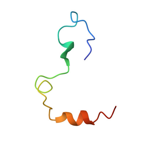
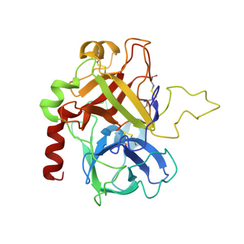
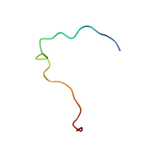
    It has been proposed that the cleaved form of protease-activated receptor 3 (PAR3) acts as a cofactor for thrombin cleavage and activation of PAR4 on murine platelets, but the molecular basis of this physiologically important effect remains elusive. X-ray crystal structures of murine thrombin bound to extracellular fragments of the murine receptors PAR3 ((38)SFNGGPQNTFEEFPLSDIE(56)) and PAR4 ((51)KSSDKPNPR downward arrow GYPGKFCANDSDTLELPASSQA(81), downward arrow = site of cleavage) have been solved at 2.0 and 3.5 A resolution, respectively. The cleaved form of PAR3, traced in the electron density maps from Gln-44 to Glu-56, makes extensive hydrophobic and electrostatic contacts with exosite I of thrombin through the fragment (47)FEEFPLSDIE(56). Occupancy of exosite I by PAR3 allosterically changes the conformation of the 60-loop and shifts the position of Trp-60d approximately 10 A with a resulting widening of the access to the active site. The PAR4 fragment, traced entirely in the electron density maps except for five C-terminal residues, clamps Trp-60d, Tyr-60a, and the aryl-binding site of thrombin with Pro-56 and Pro-58 at the P2 and P4 positions and engages the primary specificity pocket with Arg-59. The fragment then leaves the active site with Gly-60 and folds into a short helical turn that directs the backbone away from exosite I and over the autolysis loop. The structures demonstrate that thrombin activation of PAR4 may occur with exosite I available to bind cofactor molecules, like the cleaved form of PAR3, whose function is to promote substrate diffusion into the active site by allosterically changing the conformation of the 60-loop.


  • Organizational Affiliation

    Department of Biochemistry and Molecular Biophysics, Washington University School of Medicine, Box 8231, St. Louis, MO 63110, USA.


Macromolecules
Find similar proteins by:  (by identity cutoff)  |  3D Structure
Entity ID: 1
MoleculeChains Sequence LengthOrganismDetailsImage
Thrombin light chain44Mus musculusMutation(s): 0 
Gene Names: F2Cf2
UniProt & NIH Common Fund Data Resources
Find proteins for P19221 (Mus musculus)
Explore P19221 
Go to UniProtKB:  P19221
IMPC:  MGI:88380
Entity Groups  
Sequence Clusters30% Identity50% Identity70% Identity90% Identity95% Identity100% Identity
UniProt GroupP19221
Sequence Annotations
Expand
  • Reference Sequence
Find similar proteins by:  (by identity cutoff)  |  3D Structure
Entity ID: 2
MoleculeChains Sequence LengthOrganismDetailsImage
Thrombin heavy chain258Mus musculusMutation(s): 1 
Gene Names: F2Cf2
UniProt & NIH Common Fund Data Resources
Find proteins for P19221 (Mus musculus)
Explore P19221 
Go to UniProtKB:  P19221
IMPC:  MGI:88380
Entity Groups  
Sequence Clusters30% Identity50% Identity70% Identity90% Identity95% Identity100% Identity
UniProt GroupP19221
Sequence Annotations
Expand
  • Reference Sequence
Find similar proteins by:  (by identity cutoff)  |  3D Structure
Entity ID: 3
MoleculeChains Sequence LengthOrganismDetailsImage
Proteinase-activated receptor 426N/AMutation(s): 0 
UniProt
Find proteins for O88634 (Mus musculus)
Explore O88634 
Go to UniProtKB:  O88634
Entity Groups  
Sequence Clusters30% Identity50% Identity70% Identity90% Identity95% Identity100% Identity
UniProt GroupO88634
Sequence Annotations
Expand
  • Reference Sequence
Small Molecules
Ligands 1 Unique
IDChains Name / Formula / InChI Key2D Diagram3D Interactions
NAG
Query on NAG

Download Ideal Coordinates CCD File 
D [auth B],
E [auth B]
2-acetamido-2-deoxy-beta-D-glucopyranose
C8 H15 N O6
OVRNDRQMDRJTHS-FMDGEEDCSA-N
Experimental Data & Validation

Experimental Data

  • Method: X-RAY DIFFRACTION
  • Resolution: 3.50 Å
  • R-Value Free: 0.319 
  • R-Value Work: 0.306 
  • R-Value Observed: 0.306 
  • Space Group: P 65 2 2
Unit Cell:
Length ( Å )Angle ( ˚ )
a = 111.153α = 90
b = 111.153β = 90
c = 179.707γ = 120
Software Package:
Software NamePurpose
CNSrefinement
StructureStudiodata collection
HKL-2000data reduction
SCALEPACKdata scaling
MOLREPphasing

Structure Validation

View Full Validation Report


Ligand Structure Quality Assessment 


Entry History 

Deposition Data

Revision History  (Full details and data files)

  • Version 1.0: 2007-07-10
    Type: Initial release
  • Version 1.1: 2008-05-01
    Changes: Version format compliance
  • Version 1.2: 2011-07-13
    Changes: Non-polymer description, Version format compliance
  • Version 1.3: 2020-07-29
    Type: Remediation
    Reason: Carbohydrate remediation
    Changes: Data collection, Derived calculations, Structure summary
  • Version 1.4: 2021-10-20
    Changes: Database references, Derived calculations, Structure summary
  • Version 1.5: 2023-08-30
    Changes: Data collection, Refinement description