2PUV

The crystal structure of isomerase domain of glucosamine-6-phosphate synthase from Candida albicans


Experimental Data Snapshot

  • Method: X-RAY DIFFRACTION
  • Resolution: 1.90 Å
  • R-Value Free: 0.212 
  • R-Value Work: 0.171 
  • R-Value Observed: 0.172 

wwPDB Validation   3D Report Full Report


Ligand Structure Quality Assessment 


This is version 1.3 of the entry. See complete history


Literature

The Crystal and Solution Studies of Glucosamine-6-phosphate Synthase from Candida albicans

Raczynska, J.Olchowy, J.Konariev, P.V.Svergun, D.I.Milewski, S.Rypniewski, W.

(2007) J Mol Biol 372: 672-688

  • DOI: https://doi.org/10.1016/j.jmb.2007.07.002
  • Primary Citation of Related Structures:  
    2POC, 2PUT, 2PUV, 2PUW

  • PubMed Abstract: 

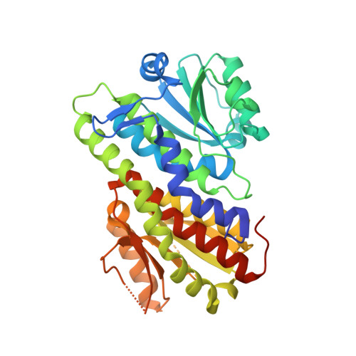
    Glucosamine 6-phosphate (GlcN-6-P) synthase is an ubiquitous enzyme that catalyses the first committed step in the reaction pathway that leads to formation of uridine 5'-diphospho-N-acetyl-D-glucosamine (UDP-GlcNAc), a precursor of macromolecules that contain amino sugars. Despite sequence similarities, the enzyme in eukaryotes is tetrameric, whereas in prokaryotes it is a dimer. The activity of eukaryotic GlcN-6-P synthase (known as Gfa1p) is regulated by feedback inhibition by UDP-GlcNAc, the end product of the reaction pathway, whereas in prokaryotes the GlcN-6-P synthase (known as GlmS) is not regulated at the post-translational level. In bacteria and fungi the enzyme is essential for cell wall synthesis. In human the enzyme is a mediator of insulin resistance. For these reasons, Gfa1p is a target in anti-fungal chemotherapy and in therapeutics for type-2 diabetes. The crystal structure of the Gfa1p isomerase domain from Candida albicans has been analysed in complex with the allosteric inhibitor UDP-GlcNAc and in the presence of glucose 6-phosphate, fructose 6-phosphate and an analogue of the reaction intermediate, 2-amino-2-deoxy-d-mannitol 6-phosphate (ADMP). A solution structure of the native Gfa1p has been deduced using small-angle X-ray scattering (SAXS). The tetrameric Gfa1p can be described as a dimer of dimers, with each half similar to the related enzyme from Escherichia coli. The core of the protein consists of the isomerase domains. UDP-GlcNAc binds, together with a metal cation, in a well-defined pocket on the surface of the isomerase domain. The residues responsible for tetramerisation and for binding UDP-GlcNAc are conserved only among eukaryotic sequences. Comparison with the previously studied GlmS from E. coli reveals differences as well as similarities in the isomerase active site. This study of Gfa1p focuses on the features that distinguish it from the prokaryotic homologue in terms of quaternary structure, control of the enzymatic activity and details of the isomerase active site.


  • Organizational Affiliation

    Institute of Bioorganic Chemistry, Polish Academy of Sciences, ul. Noskowskiego 12/14, 61-704 Poznan, Poland.


Macromolecules
Find similar proteins by:  (by identity cutoff)  |  3D Structure
Entity ID: 1
MoleculeChains Sequence LengthOrganismDetailsImage
isomerase domain of glutamine-fructose-6-phosphate transaminase (isomerizing)
A, B, C, D
367Candida albicans SC5314Mutation(s): 0 
Gene Names: GFA1
EC: 2.6.1.16
UniProt
Find proteins for P53704 (Candida albicans (strain SC5314 / ATCC MYA-2876))
Explore P53704 
Go to UniProtKB:  P53704
Entity Groups  
Sequence Clusters30% Identity50% Identity70% Identity90% Identity95% Identity100% Identity
UniProt GroupP53704
Sequence Annotations
Expand
  • Reference Sequence
Small Molecules
Ligands 4 Unique
IDChains Name / Formula / InChI Key2D Diagram3D Interactions
UD1
Query on UD1

Download Ideal Coordinates CCD File 
G [auth A],
J [auth B],
N [auth C],
Q [auth D]
URIDINE-DIPHOSPHATE-N-ACETYLGLUCOSAMINE
C17 H27 N3 O17 P2
LFTYTUAZOPRMMI-CFRASDGPSA-N
M6R
Query on M6R

Download Ideal Coordinates CCD File 
H [auth A],
K [auth B],
O [auth C],
R [auth D]
5-AMINO-5-DEOXY-1-O-PHOSPHONO-D-MANNITOL
C6 H16 N O8 P
LBNVXZROMBUNNQ-KVTDHHQDSA-N
ACT
Query on ACT

Download Ideal Coordinates CCD File 
E [auth A],
L [auth C]
ACETATE ION
C2 H3 O2
QTBSBXVTEAMEQO-UHFFFAOYSA-M
NA
Query on NA

Download Ideal Coordinates CCD File 
F [auth A],
I [auth B],
M [auth C],
P [auth D]
SODIUM ION
Na
FKNQFGJONOIPTF-UHFFFAOYSA-N
Experimental Data & Validation

Experimental Data

  • Method: X-RAY DIFFRACTION
  • Resolution: 1.90 Å
  • R-Value Free: 0.212 
  • R-Value Work: 0.171 
  • R-Value Observed: 0.172 
  • Space Group: P 1 21 1
Unit Cell:
Length ( Å )Angle ( ˚ )
a = 66.121α = 90
b = 118.167β = 91.87
c = 100.137γ = 90
Software Package:
Software NamePurpose
MAR345data collection
PHASERphasing
REFMACrefinement
DENZOdata reduction
SCALEPACKdata scaling

Structure Validation

View Full Validation Report


Ligand Structure Quality Assessment 


Entry History 

Deposition Data

Revision History  (Full details and data files)

  • Version 1.0: 2007-09-11
    Type: Initial release
  • Version 1.1: 2011-07-13
    Changes: Source and taxonomy, Version format compliance
  • Version 1.2: 2017-10-18
    Changes: Refinement description
  • Version 1.3: 2023-08-30
    Changes: Data collection, Database references, Derived calculations, Refinement description