2PMN

Crystal structure of PfPK7 in complex with an ATP-site inhibitor


Experimental Data Snapshot

  • Method: X-RAY DIFFRACTION
  • Resolution: 2.80 Å
  • R-Value Free: 0.296 
  • R-Value Work: 0.219 
  • R-Value Observed: 0.223 

wwPDB Validation   3D Report Full Report


Ligand Structure Quality Assessment 


This is version 1.2 of the entry. See complete history


Literature

Structures of P. falciparum protein kinase 7 identify an activation motif and leads for inhibitor design.

Merckx, A.Echalier, A.Langford, K.Sicard, A.Langsley, G.Joore, J.Doerig, C.Noble, M.Endicott, J.

(2008) Structure 16: 228-238

  • DOI: https://doi.org/10.1016/j.str.2007.11.014
  • Primary Citation of Related Structures:  
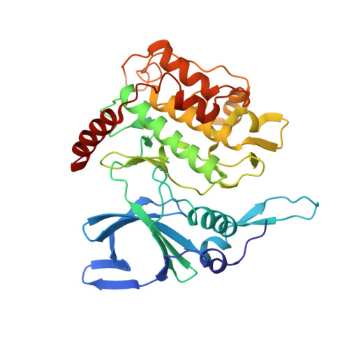
    2PMN, 2PMO

  • PubMed Abstract: 

    Malaria is a major threat to world health. The identification of parasite targets for drug development is a priority and parasitic protein kinases suggest themselves as suitable targets as many display profound structural and functional divergences from their host counterparts. In this paper, we describe the structure of the orphan protein kinase, Plasmodium falciparum protein kinase 7 (PFPK7). Several Plasmodium protein kinases contain extensive insertions, and the structure of PFPK7 reveals how these may be accommodated as excursions from the canonical eukaryotic protein kinase fold. The constitutively active conformation of PFPK7 is stabilized by a structural motif in which the role of the conserved phosphorylated residue that assists in structuring the activation loop of many protein kinases is played by an arginine residue. We identify two series of PFPK7 ATP-competitive inhibitors and suggest further developments for the design of selective and potent PFPK7 lead compounds as potential antimalarials.


  • Organizational Affiliation

    The Laboratory of Molecular Biophysics, Department of Biochemistry, University of Oxford, South Parks Road, Oxford OX1 3QU, United Kingdom.


Macromolecules
Find similar proteins by:  (by identity cutoff)  |  3D Structure
Entity ID: 1
MoleculeChains Sequence LengthOrganismDetailsImage
Ser/Thr protein kinase, putativeA [auth X]348Plasmodium falciparumMutation(s): 0 
Gene Names: PFB0605w
UniProt
Find proteins for Q7YTF7 (Plasmodium falciparum (isolate 3D7))
Explore Q7YTF7 
Go to UniProtKB:  Q7YTF7
Entity Groups  
Sequence Clusters30% Identity50% Identity70% Identity90% Identity95% Identity100% Identity
UniProt GroupQ7YTF7
Sequence Annotations
Expand
  • Reference Sequence
Small Molecules
Ligands 1 Unique
IDChains Name / Formula / InChI Key2D Diagram3D Interactions
K51
Query on K51

Download Ideal Coordinates CCD File 
B [auth X]4-(6-{[(1S)-1-(HYDROXYMETHYL)-2-METHYLPROPYL]AMINO}IMIDAZO[1,2-B]PYRIDAZIN-3-YL)BENZONITRILE
C18 H19 N5 O
CQZVJEKKNHOFNB-OAHLLOKOSA-N
Binding Affinity Annotations 
IDSourceBinding Affinity
K51 PDBBind:  2PMN IC50: 6300 (nM) from 1 assay(s)
BindingDB:  2PMN IC50: 280 (nM) from 1 assay(s)
Experimental Data & Validation

Experimental Data

  • Method: X-RAY DIFFRACTION
  • Resolution: 2.80 Å
  • R-Value Free: 0.296 
  • R-Value Work: 0.219 
  • R-Value Observed: 0.223 
  • Space Group: C 2 2 21
Unit Cell:
Length ( Å )Angle ( ˚ )
a = 73.406α = 90
b = 82.084β = 90
c = 138.962γ = 90
Software Package:
Software NamePurpose
REFMACrefinement
DNAdata collection
MOSFLMdata reduction
SCALAdata scaling
MOLREPphasing

Structure Validation

View Full Validation Report


Ligand Structure Quality Assessment 


Entry History 

Deposition Data

Revision History  (Full details and data files)

  • Version 1.0: 2008-01-22
    Type: Initial release
  • Version 1.1: 2011-07-13
    Changes: Advisory, Version format compliance
  • Version 1.2: 2023-08-30
    Changes: Data collection, Database references, Derived calculations, Refinement description