2PGI

THE CRYSTAL STRUCTURE OF PHOSPHOGLUCOSE ISOMERASE-AN ENZYME WITH AUTOCRINE MOTILITY FACTOR ACTIVITY IN TUMOR CELLS


Experimental Data Snapshot

  • Method: X-RAY DIFFRACTION
  • Resolution: 2.30 Å
  • R-Value Free: 0.278 
  • R-Value Work: 0.185 
  • R-Value Observed: 0.185 

wwPDB Validation   3D Report Full Report


This is version 1.3 of the entry. See complete history


Literature

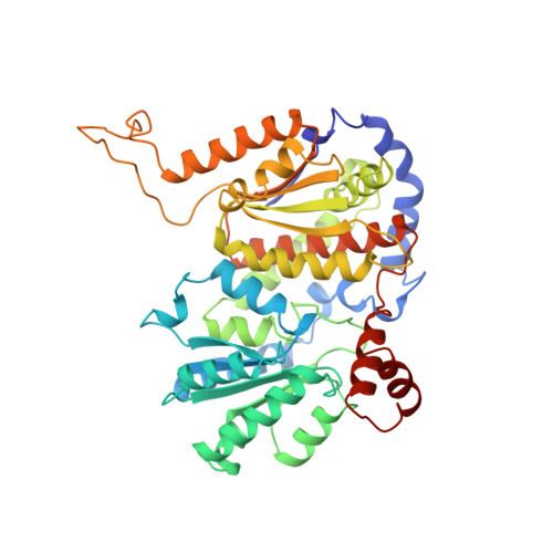
The crystal structure of a multifunctional protein: phosphoglucose isomerase/autocrine motility factor/neuroleukin.

Sun, Y.J.Chou, C.C.Chen, W.S.Wu, R.T.Meng, M.Hsiao, C.D.

(1999) Proc Natl Acad Sci U S A 96: 5412-5417

  • DOI: https://doi.org/10.1073/pnas.96.10.5412
  • Primary Citation of Related Structures:  
    2PGI

  • PubMed Abstract: 

    Phosphoglucose isomerase (PGI) plays a central role in both the glycolysis and the gluconeogenesis pathways. We present here the complete crystal structure of PGI from Bacillus stearothermophilus at 2.3-A resolution. We show that PGI has cell-motility-stimulating activity on mouse colon cancer cells similar to that of endogenous autocrine motility factor (AMF). PGI can also enhance neurite outgrowth on neuronal progenitor cells similar to that observed for neuroleukin. The results confirm that PGI is neuroleukin and AMF. PGI has an open twisted alpha/beta structural motif consisting of two globular domains and two protruding parts. Based on this substrate-free structure, together with the previously published biological, biochemical, and modeling results, we postulate a possible substrate-binding site that is located within the domains' interface for PGI and AMF. In addition, the structure provides evidence suggesting that the top part of the large domain together with one of the protruding loops might participate in inducing the neurotrophic activity.


  • Organizational Affiliation

    Institute of Molecular Biology, Academia Sinica, Taipei, Taiwan 11529, Republic of China.


Macromolecules
Find similar proteins by:  (by identity cutoff)  |  3D Structure
Entity ID: 1
MoleculeChains Sequence LengthOrganismDetailsImage
PHOSPHOGLUCOSE ISOMERASE445Geobacillus stearothermophilusMutation(s): 0 
Gene Names: PGIB
EC: 5.3.1.9
UniProt
Find proteins for P13376 (Geobacillus stearothermophilus)
Explore P13376 
Go to UniProtKB:  P13376
Entity Groups  
Sequence Clusters30% Identity50% Identity70% Identity90% Identity95% Identity100% Identity
UniProt GroupP13376
Sequence Annotations
Expand
  • Reference Sequence
Experimental Data & Validation

Experimental Data

  • Method: X-RAY DIFFRACTION
  • Resolution: 2.30 Å
  • R-Value Free: 0.278 
  • R-Value Work: 0.185 
  • R-Value Observed: 0.185 
  • Space Group: I 2 2 2
Unit Cell:
Length ( Å )Angle ( ˚ )
a = 75.1α = 90
b = 93.73β = 90
c = 171.9γ = 90
Software Package:
Software NamePurpose
X-PLORmodel building
X-PLORrefinement
DENZOdata reduction
SCALEPACKdata scaling
X-PLORphasing

Structure Validation

View Full Validation Report


Entry History 

Deposition Data

Revision History  (Full details and data files)

  • Version 1.0: 1999-06-15
    Type: Initial release
  • Version 1.1: 2008-03-24
    Changes: Version format compliance
  • Version 1.2: 2011-07-13
    Changes: Derived calculations, Version format compliance
  • Version 1.3: 2024-02-21
    Changes: Data collection, Database references, Other