2OMB

Bence Jones KWR Protein- Immunoglobulin Light Chain Dimer, P3(1)21 Crystal Form


Experimental Data Snapshot

  • Method: X-RAY DIFFRACTION
  • Resolution: 2.90 Å
  • R-Value Free: 0.284 
  • R-Value Work: 0.235 
  • R-Value Observed: 0.238 

wwPDB Validation   3D Report Full Report


This is version 2.1 of the entry. See complete history


Literature

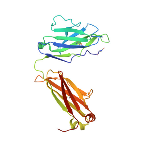
Bence Jones KWR protein structures determined by X-ray crystallography.

Makino, D.L.Henschen-Edman, A.H.Larson, S.B.McPherson, A.

(2007) Acta Crystallogr D Biol Crystallogr 63: 780-792

  • DOI: https://doi.org/10.1107/S0907444907021981
  • Primary Citation of Related Structures:  
    2OLD, 2OMB, 2OMN

  • PubMed Abstract: 

    A Bence Jones protein isolated in the early 1960s from a patient (initials KWR) suffering from plasma-cell dyscrasia was crystallized and its structure was analyzed in four different unit cells by X-ray diffraction. The final models of the molecule in all crystal forms were virtually the same, although the elbow angles relating the constant and variable domains of the Bence Jones dimers varied over a range of 10 degrees. The tetragonal form had an R factor of 22.6% and an R(free) of 28.3% at 2.2 A resolution. Phosphate or sulfate ions (depending on the crystallization conditions) were found in the antigen-combining sites in all crystals, as well as an unidentified ligand tightly bound in the hydrophobic 'deep pocket' beneath the antigen-binding site. The ligand was treated as a phenol molecule. Two trigonal crystal forms were among those solved. One was grown at pH 4.0 and the other was only obtained after sitting for more than eight months at room temperature. The latter crystal was composed of molecules that were degraded in their constant domains. Both low pH and proteolytic degradation of constant domains are known to promote the polymerization of some Bence Jones proteins into amyloid fibrils. Indeed, in both trigonal crystal forms the molecules are organized with pseudo-hexagonal symmetry about the unique crystallographic axes in a manner suggestive of such fibrils. The arrangement of Bence Jones dimers is also consistent with other observations regarding Bence Jones amyloid-fibril structure and current models.


  • Organizational Affiliation

    Department of Molecular Biology and Biochemistry, University of California, Irvine, CA 92697-3900, USA.


Macromolecules
Find similar proteins by:  (by identity cutoff)  |  3D Structure
Entity ID: 1
MoleculeChains Sequence LengthOrganismDetailsImage
Bence Jones KWR Protein - Immunoglobulin Light Chain
A, B, C, D
217Homo sapiensMutation(s): 0 
Entity Groups  
Sequence Clusters30% Identity50% Identity70% Identity90% Identity95% Identity100% Identity
Sequence Annotations
Expand
  • Reference Sequence
Small Molecules
Ligands 2 Unique
IDChains Name / Formula / InChI Key2D Diagram3D Interactions
SO4
Query on SO4

Download Ideal Coordinates CCD File 
E [auth A]
F [auth A]
G [auth A]
I [auth B]
J [auth B]
E [auth A],
F [auth A],
G [auth A],
I [auth B],
J [auth B],
K [auth C],
L [auth C],
M [auth D],
N [auth D]
SULFATE ION
O4 S
QAOWNCQODCNURD-UHFFFAOYSA-L
IPH
Query on IPH

Download Ideal Coordinates CCD File 
H [auth A],
O [auth D]
PHENOL
C6 H6 O
ISWSIDIOOBJBQZ-UHFFFAOYSA-N
Modified Residues  1 Unique
IDChains TypeFormula2D DiagramParent
PCA
Query on PCA
A, B, C, D
L-PEPTIDE LINKINGC5 H7 N O3GLN
Experimental Data & Validation

Experimental Data

  • Method: X-RAY DIFFRACTION
  • Resolution: 2.90 Å
  • R-Value Free: 0.284 
  • R-Value Work: 0.235 
  • R-Value Observed: 0.238 
  • Space Group: P 31 2 1
Unit Cell:
Length ( Å )Angle ( ˚ )
a = 153.52α = 90
b = 153.52β = 90
c = 93.95γ = 120
Software Package:
Software NamePurpose
CNSrefinement
MAR345data collection
HKL-2000data reduction
HKL-2000data scaling
CNSphasing

Structure Validation

View Full Validation Report


Entry History 

Deposition Data

Revision History  (Full details and data files)

  • Version 1.0: 2007-07-17
    Type: Initial release
  • Version 1.1: 2008-05-01
    Changes: Version format compliance
  • Version 1.2: 2011-07-13
    Changes: Version format compliance
  • Version 2.0: 2019-12-25
    Changes: Derived calculations, Polymer sequence
  • Version 2.1: 2023-08-30
    Changes: Data collection, Database references, Derived calculations, Refinement description