Multifaceted recognition of vertebrate Rev1 by translesion polymerases zeta and kappa.
Wojtaszek, J., Liu, J., D'Souza, S., Wang, S., Xue, Y., Walker, G.C., Zhou, P.(2012) J Biol Chem 287: 26400-26408
- PubMed: 22700975
- DOI: https://doi.org/10.1074/jbc.M112.380998
- Primary Citation of Related Structures:
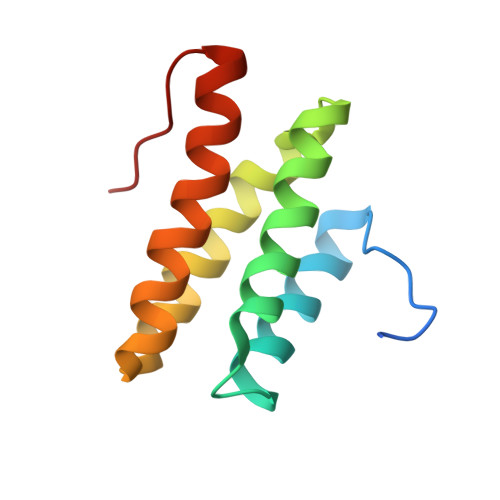
2LSG, 2LSJ - PubMed Abstract:
Translesion synthesis is a fundamental biological process that enables DNA replication across lesion sites to ensure timely duplication of genetic information at the cost of replication fidelity, and it is implicated in development of cancer drug resistance after chemotherapy. The eukaryotic Y-family polymerase Rev1 is an essential scaffolding protein in translesion synthesis. Its C-terminal domain (CTD), which interacts with translesion polymerase ζ through the Rev7 subunit and with polymerases κ, ι, and η in vertebrates through the Rev1-interacting region (RIR), is absolutely required for function. We report the first solution structures of the mouse Rev1 CTD and its complex with the Pol κ RIR, revealing an atypical four-helix bundle. Using yeast two-hybrid assays, we have identified a Rev7-binding surface centered at the α2-α3 loop and N-terminal half of α3 of the Rev1 CTD. Binding of the mouse Pol κ RIR to the Rev1 CTD induces folding of the disordered RIR peptide into a three-turn α-helix, with the helix stabilized by an N-terminal cap. RIR binding also induces folding of a disordered N-terminal loop of the Rev1 CTD into a β-hairpin that projects over the shallow α1-α2 surface and creates a deep hydrophobic cavity to interact with the essential FF residues juxtaposed on the same side of the RIR helix. Our combined structural and biochemical studies reveal two distinct surfaces of the Rev1 CTD that separately mediate the assembly of extension and insertion translesion polymerase complexes and provide a molecular framework for developing novel cancer therapeutics to inhibit translesion synthesis.
Organizational Affiliation:
Department of Biochemistry, Duke University, Medical Center, Durham, North Carolina 27710, USA.