Structural Basis for 5'-End-Specific Recognition of Single-Stranded DNA by the R3H Domain from Human Smubp-2
Jaudzems, K., Jia, X., Yagi, H., Zhulenkovs, D., Graham, B., Otting, G., Liepinsh, E.(2012) J Mol Biol 12: 760-767
- PubMed: 22999958
- DOI: https://doi.org/10.1016/j.jmb.2012.09.010
- Primary Citation of Related Structures:
2LRR - PubMed Abstract:
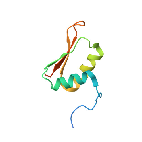
The R3H domain is a conserved sequence motif in nucleic acid binding proteins. Previously, we reported the solution structure of the R3H domain and identified a putative nucleic acid binding site composed of three conserved basic residues [Liepinsh, E., Leonchiks, A., Sharipo, A., Guignard, L. & Otting, G. (2003). Solution structure of the R3H domain from human Sμbp-2. J. Mol. Biol.326, 217-223]. Here, we determine the binding affinities of mononucleotides and dinucleotides for the R3H domain from human Sμbp-2 (Sμbp2-R3H) and map their binding sites on the protein's surface. Although the binding affinities show up to 260-fold selectivity between different nucleotides, their binding sites and conformations seem very similar. Further, we report the NMR structure of the Sμbp2-R3H in complex with deoxyguanosine 5'-monophosphate (dGMP) mimicking the 5'-end of single-stranded DNA. Pseudocontact shifts from a paramagnetic lanthanide tag attached to residue 731 in the mutant A731C confirmed that binding of dGMP brings a loop of the protein into closer proximity. The structure provides the first structural insight into single-stranded nucleic acid recognition by the R3H domain and shows that the R3H domain specifically binds the phosphorylated 5'-end through electrostatic interactions with the two conserved arginines and stacking interactions with the highly conserved histidine.
Organizational Affiliation:
Department of Physical Organic Chemistry, Latvian Institute of Organic Synthesis, Latvia. kristaps.jaudzems@osi.lv