2LIG

THREE-DIMENSIONAL STRUCTURES OF THE LIGAND-BINDING DOMAIN OF THE BACTERIAL ASPARTATE RECEPTOR WITH AND WITHOUT A LIGAND


Experimental Data Snapshot

  • Method: X-RAY DIFFRACTION
  • Resolution: 2.00 Å
  • R-Value Work: 0.176 
  • R-Value Observed: 0.176 

wwPDB Validation   3D Report Full Report


This is version 1.3 of the entry. See complete history


Literature

Three-dimensional structures of the ligand-binding domain of the bacterial aspartate receptor with and without a ligand.

Milburn, M.V.Prive, G.G.Milligan, D.L.Scott, W.G.Yeh, J.Jancarik, J.Koshland Jr., D.E.Kim, S.H.

(1991) Science 254: 1342-1347

  • DOI: https://doi.org/10.1126/science.1660187
  • Primary Citation of Related Structures:  
    1LIH, 2LIG

  • PubMed Abstract: 

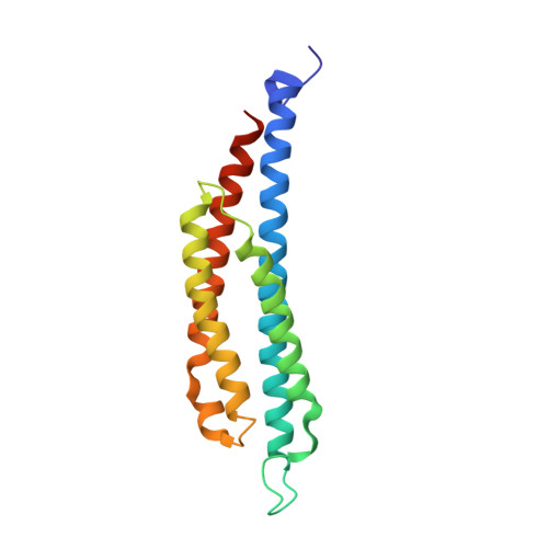
    The three-dimensional structure of an active, disulfide cross-linked dimer of the ligand-binding domain of the Salmonella typhimurium aspartate receptor and that of an aspartate complex have been determined by x-ray crystallographic methods at 2.4 and 2.0 angstrom (A) resolution, respectively. A single subunit is a four-alpha-helix bundle with two long amino-terminal and carboxyl-terminal helices and two shorter helices that form a cylinder 20 A in diameter and more than 70 A long. The two subunits in the disulfide-bonded dimer are related by a crystallographic twofold axis in the apo structure, but by a noncrystallographic twofold axis in the aspartate complex structure. The latter structure reveals that the ligand binding site is located more than 60 A from the presumed membrane surface and is at the interface of the two subunits. Aspartate binds between two alpha helices from one subunit and one alpha helix from the other in a highly charged pocket formed by three arginines. The comparison of the apo and aspartate complex structures shows only small structural changes in the individual subunits, except for one loop region that is disordered, but the subunits appear to change orientation relative to each other. The structures of the two forms of this protein provide a step toward understanding the mechanisms of transmembrane signaling.


  • Organizational Affiliation

    Department of Chemistry, University of California, Berkeley 94720.


Macromolecules
Find similar proteins by:  (by identity cutoff)  |  3D Structure
Entity ID: 1
MoleculeChains Sequence LengthOrganismDetailsImage
ASPARTATE RECEPTOR
A, B
164Salmonella enterica subsp. enterica serovar TyphimuriumMutation(s): 0 
UniProt
Find proteins for P02941 (Salmonella typhimurium (strain LT2 / SGSC1412 / ATCC 700720))
Explore P02941 
Go to UniProtKB:  P02941
Entity Groups  
Sequence Clusters30% Identity50% Identity70% Identity90% Identity95% Identity100% Identity
UniProt GroupP02941
Sequence Annotations
Expand
  • Reference Sequence
Small Molecules
Ligands 3 Unique
IDChains Name / Formula / InChI Key2D Diagram3D Interactions
PHN
Query on PHN

Download Ideal Coordinates CCD File 
F [auth A]1,10-PHENANTHROLINE
C12 H8 N2
DGEZNRSVGBDHLK-UHFFFAOYSA-N
ASP
Query on ASP

Download Ideal Coordinates CCD File 
E [auth A]ASPARTIC ACID
C4 H7 N O4
CKLJMWTZIZZHCS-REOHCLBHSA-N
SO4
Query on SO4

Download Ideal Coordinates CCD File 
C [auth A],
D [auth A]
SULFATE ION
O4 S
QAOWNCQODCNURD-UHFFFAOYSA-L
Experimental Data & Validation

Experimental Data

  • Method: X-RAY DIFFRACTION
  • Resolution: 2.00 Å
  • R-Value Work: 0.176 
  • R-Value Observed: 0.176 
  • Space Group: P 32 2 1
Unit Cell:
Length ( Å )Angle ( ˚ )
a = 80.38α = 90
b = 80.38β = 90
c = 91.35γ = 120
Software Package:
Software NamePurpose
X-PLORmodel building
X-PLORrefinement
X-PLORphasing

Structure Validation

View Full Validation Report


Entry History 

Deposition Data

Revision History  (Full details and data files)

  • Version 1.0: 1995-09-15
    Type: Initial release
  • Version 1.1: 2008-03-03
    Changes: Version format compliance
  • Version 1.2: 2011-07-13
    Changes: Version format compliance
  • Version 1.3: 2017-11-29
    Changes: Derived calculations, Other