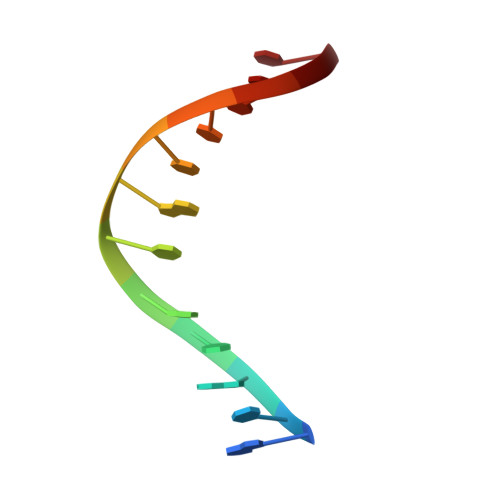
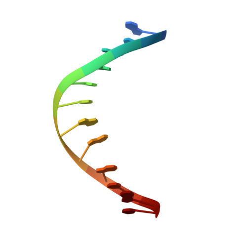
Structures of (5'S)-8,5'-Cyclo-2'-deoxyguanosine Mismatched with dA or dT.
Huang, H., Das, R.S., Basu, A.K., Stone, M.P.(2012) Chem Res Toxicol 25: 478-490
- PubMed: 22309170
- DOI: https://doi.org/10.1021/tx2005053
- Primary Citation of Related Structures:
2LFX, 2LFY - PubMed Abstract:
Diastereomeric 8,5'-cyclopurine 2'-deoxynucleosides, containing a covalent bond between the deoxyribose and the purine base, are induced in DNA by ionizing radiation. They are suspected to play a role in the etiology of neurodegeneration in xeroderma pigmentosum patients. If not repaired, the S-8,5'-cyclo-2'-deoxyguanosine lesion (S-cdG) induces Pol V-dependent mutations at a frequency of 34% in Escherichia coli. Most are S-cdG → A transitions, suggesting mis-incorporation of dTTP opposite the lesion during replication bypass, although low levels of S-cdG → T transversions, arising from mis-incorporation of dATP, are also observed. We report the structures of 5'-d(GTGCXTGTTTGT)-3'·5'-d(ACAAACAYGCAC)-3', where X denotes S-cdG and Y denotes either dA or dT, corresponding to the situation following mis-insertion of either dTTP or dATP opposite the S-cdG lesion. The S-cdG·dT mismatch pair adopts a wobble base pairing. This provides a plausible rationale for the S-cdG → A transitions. The S-cdG·dA mismatch pair differs in conformation from the dG·dA mismatch pair. For the S-cdG·dA mismatch pair, both S-cdG and dA intercalate, but no hydrogen bonding is observed between S-cdG and dA. This is consistent with the lower levels of S-cdG → T transitions in E. coli.
Organizational Affiliation:
Department of Chemistry, Center in Molecular Toxicology, and Center for Structural Biology, Vanderbilt-Ingram Cancer Center, Vanderbilt University , Nashville, Tennessee 37235, United States.