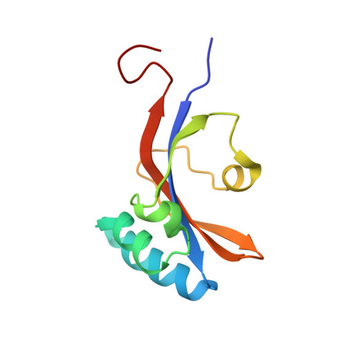
The prolyl isomerase domain of PpiD from Escherichia coli shows a parvulin fold but is devoid of catalytic activity.
Weininger, U., Jakob, R.P., Kovermann, M., Balbach, J., Schmid, F.X.(2009) Protein Sci 19: 6-18
- PubMed: 19866485
- DOI: https://doi.org/10.1002/pro.277
- Primary Citation of Related Structures:
2KGJ - PubMed Abstract:
PpiD is a periplasmic folding helper protein of Escherichia coli. It consists of an N-terminal helix that anchors PpiD in the inner membrane near the SecYEG translocon, followed by three periplasmic domains. The second domain (residues 264-357) shows homology to parvulin-like prolyl isomerases. This domain is a well folded, stable protein and follows a simple two-state folding mechanism. In its solution structure, as determined by NMR spectroscopy, it resembles most closely the first parvulin domain of the SurA protein, which resides in the periplasm of E. coli as well. A previously reported prolyl isomerase activity of PpiD could not be reproduced when using improved protease-free peptide assays or assays with refolding proteins as substrates. The parvulin domain of PpiD interacts, however, with a proline-containing tetrapeptide, and the binding site, as identified by NMR resonance shift analysis, colocalized with the catalytic sites of other parvulins. In its structure, the parvulin domain of PpiD resembles most closely the inactive first parvulin domain of SurA, which is part of the chaperone unit of this protein and presumably involved in substrate recognition.
Organizational Affiliation:
Institut für Physik, Biophysik, and Mitteldeutsches Zentrum für Struktur und Dynamik der Proteine (MZP), Martin-Luther-Universität Halle-Wittenberg, D-06120 Halle(Saale), Germany.