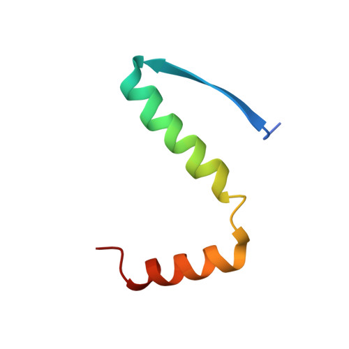
Conformational stability and activity of p73 require a second helix in the tetramerization domain.
Coutandin, D., Lohr, F., Niesen, F.H., Ikeya, T., Weber, T.A., Schafer, B., Zielonka, E.M., Bullock, A.N., Yang, A., Guntert, P., Knapp, S., McKeon, F., Ou, H.D., Dotsch, V.(2009) Cell Death Differ 16: 1582-1589
- PubMed: 19763140
- DOI: https://doi.org/10.1038/cdd.2009.139
- Primary Citation of Related Structures:
2KBY - PubMed Abstract:
p73 and p63, the two ancestral members of the p53 family, are involved in neurogenesis, epithelial stem cell maintenance and quality control of female germ cells. The highly conserved oligomerization domain (OD) of tumor suppressor p53 is essential for its biological functions, and its structure was believed to be the prototype for all three proteins. However, we report that the ODs of p73 and p63 differ from the OD of p53 by containing an additional alpha-helix that is not present in the structure of the p53 OD. Deletion of this helix causes a dissociation of the OD into dimers; it also causes conformational instability and reduces the transcriptional activity of p73. Moreover, we show that ODs of p73 and p63 strongly interact and that a large number of different heterotetramers are supported by the additional helix. Detailed analysis shows that the heterotetramer consisting of two homodimers is thermodynamically more stable than the two homotetramers. No heterooligomerization between p53 and the p73/p63 subfamily was observed, supporting the notion of functional orthogonality within the p53 family.
Organizational Affiliation:
Institute of Biophysical Chemistry and Center for Biomolecular Magnetic Resonance and Cluster of Excellence Macromolecular Complexes (CEF), Goethe University, Frankfurt/Main, Germany.