2JJF

N328A mutant of M. tuberculosis Rv3290c


Experimental Data Snapshot

  • Method: X-RAY DIFFRACTION
  • Resolution: 1.95 Å
  • R-Value Free: 0.185 
  • R-Value Work: 0.166 
  • R-Value Observed: 0.167 

wwPDB Validation   3D Report Full Report


This is version 1.1 of the entry. See complete history


Literature

Mutational Analysis of Mycobacterium Tuberculosis Lysine Epsilon-Aminotransferase and Inhibitor Co-Crystal Structures, Reveals Distinct Binding Modes.

Tripathi, S.M.Agarwal, A.Ramachandran, R.

(2015) Biochem Biophys Res Commun 463: 154

  • DOI: https://doi.org/10.1016/j.bbrc.2015.05.055
  • Primary Citation of Related Structures:  
    2JJE, 2JJF, 2JJG, 2JJH

  • PubMed Abstract: 

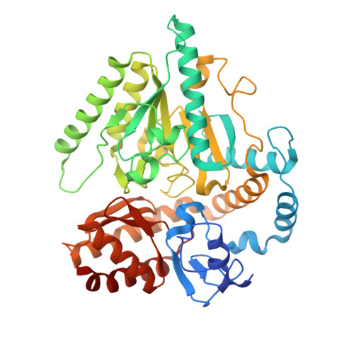
    Lysine ɛ-aminotransferase (LAT) converts lysine to α-aminoadipate-δ-semialdehyde in a PLP-mediated reaction. We mutated active-site T330, N328 and E243, and structurally rationalized their properties. T330A and T330S mutants cannot bind PLP and are inactive. N328A although inactive, binds to PLP. E243A retains activity, but binds α-ketoglutarate in a different conformation. We had earlier identified 2-aminomethyl piperidine derivative as a LAT inhibitor. The co-crystal structure reveals that it mimics binding of C5 substrates and exhibits two binding modes. E243, that shields R422 in the apo enzyme, exhibits conformational changes to permit the binding of the inhibitor in one of the binding modes. Structure-based analysis of bound water in the active site suggests optimization strategies for synthesis of improved inhibitors.


  • Organizational Affiliation

    Molecular & Structural Biology Division, Central Drug Research Institute, Sector 10, Jankipuram Extension, Sitapur Road, Lucknow 226031, U.P., India.


Macromolecules
Find similar proteins by:  (by identity cutoff)  |  3D Structure
Entity ID: 1
MoleculeChains Sequence LengthOrganismDetailsImage
L-LYSINE EPSILON AMINOTRANSFERASE449Mycobacterium tuberculosisMutation(s): 1 
EC: 2.6.1.36
UniProt
Find proteins for P9WQ77 (Mycobacterium tuberculosis (strain ATCC 25618 / H37Rv))
Explore P9WQ77 
Go to UniProtKB:  P9WQ77
Entity Groups  
Sequence Clusters30% Identity50% Identity70% Identity90% Identity95% Identity100% Identity
UniProt GroupP9WQ77
Sequence Annotations
Expand
  • Reference Sequence
Experimental Data & Validation

Experimental Data

  • Method: X-RAY DIFFRACTION
  • Resolution: 1.95 Å
  • R-Value Free: 0.185 
  • R-Value Work: 0.166 
  • R-Value Observed: 0.167 
  • Space Group: P 31 2 1
Unit Cell:
Length ( Å )Angle ( ˚ )
a = 103.407α = 90
b = 103.407β = 90
c = 98.162γ = 120
Software Package:
Software NamePurpose
REFMACrefinement

Structure Validation

View Full Validation Report


Entry History 

Deposition Data

Revision History  (Full details and data files)

  • Version 1.0: 2009-06-30
    Type: Initial release
  • Version 1.1: 2017-01-25
    Changes: Database references, Derived calculations, Other, Structure summary, Version format compliance