2JFE

The crystal structure of human cytosolic beta-glucosidase


Experimental Data Snapshot

  • Method: X-RAY DIFFRACTION
  • Resolution: 2.70 Å
  • R-Value Free: 0.280 
  • R-Value Work: 0.197 
  • R-Value Observed: 0.201 

wwPDB Validation   3D Report Full Report


This is version 2.1 of the entry. See complete history


Literature

The Crystal Structure of Human Cytosolic Beta-Glucosidase Unravels the Substrate Aglycone Specificity of a Family 1 Glycoside Hydrolase

Tribolo, S.Berrin, J.G.Kroon, P.A.Czjzek, M.Juge, N.

(2007) J Mol Biol 370: 964

  • DOI: https://doi.org/10.1016/j.jmb.2007.05.034
  • Primary Citation of Related Structures:  
    2JFE

  • PubMed Abstract: 

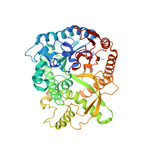
    Human cytosolic beta-glucosidase (hCBG) is a xenobiotic-metabolizing enzyme that hydrolyses certain flavonoid glucosides, with specificity depending on the aglycone moiety, the type of sugar and the linkage between them. In this study, the substrate preference of this enzyme was investigated by mutational analysis, X-ray crystallography and homology modelling. The crystal structure of hCBG was solved by the molecular replacement method and refined at 2.7 A resolution. The main-chain fold of the enzyme belongs to the (beta/alpha)(8) barrel structure, which is common to family 1 glycoside hydrolases. The active site is located at the bottom of a pocket (about 16 A deep) formed by large surface loops, surrounding the C termini of the barrel of beta-strands. As for all the clan of GH-A enzymes, the two catalytic glutamate residues are located on strand 4 (the acid/base Glu165) and on strand 7 (the nucleophile Glu373). Although many features of hCBG were shown to be very similar to previously described enzymes from this family, crucial differences were observed in the surface loops surrounding the aglycone binding site, and these are likely to strongly influence the substrate specificity. The positioning of a substrate molecule (quercetin-4'-glucoside) by homology modelling revealed that hydrophobic interactions dominate the binding of the aglycone moiety. In particular, Val168, Trp345, Phe225, Phe179, Phe334 and Phe433 were identified as likely to be important in determining substrate specificity in hCBG, and site-directed mutagenesis supported a key role for some of these residues.


  • Organizational Affiliation

    Institute of Food Research, Norwich Research Park, Colney, Norwich NR4 7UA, UK.


Macromolecules
Find similar proteins by:  (by identity cutoff)  |  3D Structure
Entity ID: 1
MoleculeChains Sequence LengthOrganismDetailsImage
CYTOSOLIC BETA-GLUCOSIDASEA [auth X]469Homo sapiensMutation(s): 0 
EC: 3.2.1.21
UniProt & NIH Common Fund Data Resources
Find proteins for Q9H227 (Homo sapiens)
Explore Q9H227 
Go to UniProtKB:  Q9H227
PHAROS:  Q9H227
Entity Groups  
Sequence Clusters30% Identity50% Identity70% Identity90% Identity95% Identity100% Identity
UniProt GroupQ9H227
Sequence Annotations
Expand
  • Reference Sequence
Oligosaccharides

Help

Entity ID: 2
MoleculeChains Length2D Diagram Glycosylation3D Interactions
2-acetamido-2-deoxy-beta-D-glucopyranose-(1-4)-2-acetamido-2-deoxy-beta-D-glucopyranoseB [auth A]2N-Glycosylation
Glycosylation Resources
GlyTouCan:  G42666HT
GlyCosmos:  G42666HT
GlyGen:  G42666HT
Experimental Data & Validation

Experimental Data

  • Method: X-RAY DIFFRACTION
  • Resolution: 2.70 Å
  • R-Value Free: 0.280 
  • R-Value Work: 0.197 
  • R-Value Observed: 0.201 
  • Space Group: P 31 2 1
Unit Cell:
Length ( Å )Angle ( ˚ )
a = 90.479α = 90
b = 90.479β = 90
c = 127.925γ = 120
Software Package:
Software NamePurpose
REFMACrefinement
MOSFLMdata reduction
SCALAdata scaling
AMoREphasing

Structure Validation

View Full Validation Report


Entry History 

Deposition Data

Revision History  (Full details and data files)

  • Version 1.0: 2007-06-19
    Type: Initial release
  • Version 1.1: 2011-05-08
    Changes: Version format compliance
  • Version 1.2: 2011-07-13
    Changes: Version format compliance
  • Version 2.0: 2020-07-29
    Type: Remediation
    Reason: Carbohydrate remediation
    Changes: Atomic model, Data collection, Derived calculations, Other, Structure summary
  • Version 2.1: 2023-12-13
    Changes: Data collection, Database references, Refinement description, Structure summary