Solution Structure of the A4 Domain of Factor Xi Sheds Light on the Mechanism of Zymogen Activation.
Samuel, D., Cheng, H., Riley, P.W., Canutescu, A.A., Nagaswami, C., Weisel, J.W., Bu, Z., Walsh, P.N., Roder, H.(2007) Proc Natl Acad Sci U S A 104: 15693
- PubMed: 17884987
- DOI: https://doi.org/10.1073/pnas.0703080104
- Primary Citation of Related Structures:
2J8J, 2J8L - PubMed Abstract:
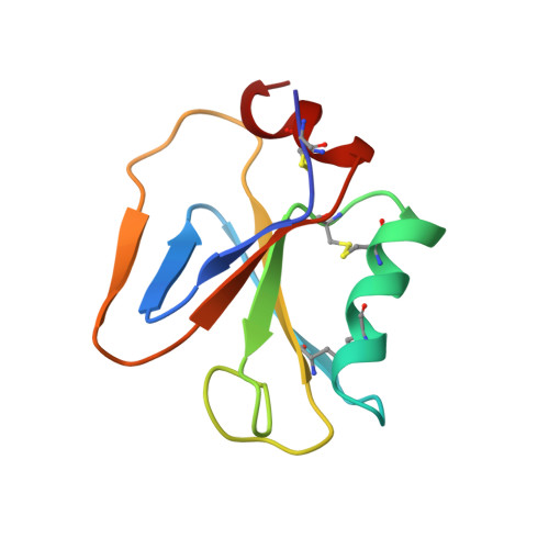
Factor XI (FXI) is a homodimeric blood coagulation protein. Each monomer comprises four tandem apple-domain repeats (A1-A4) and a serine protease domain. We report here the NMR solution structure of the A4 domain (residues 272-361), which mediates formation of the disulfide-linked FXI dimer. A4 exhibits characteristic features of the plasminogen apple nematode domain family, including a five-stranded beta-sheet flanked by an alpha-helix on one side and a two-stranded beta-sheet on the other. In addition, the solution structure reveals a second alpha-helix at the C terminus. Comparison with a recent crystal structure of full-length FXI, combined with molecular modeling, suggests that the C-terminal helix is formed only upon proteolytic activation. The newly formed helix disrupts interdomain contacts and reorients the catalytic domains, bringing the active sites into close proximity. This hypothesis is supported by small-angle x-ray scattering and electron microscopy data, which indicate that FXI activation is accompanied by a major change in shape. The results are consistent with biochemical evidence that activated FXI cleaves its substrate at two positions without release of an intermediate.
Organizational Affiliation:
Division of Basic Science, Fox Chase Cancer Center, Philadelphia, PA 19111, USA.