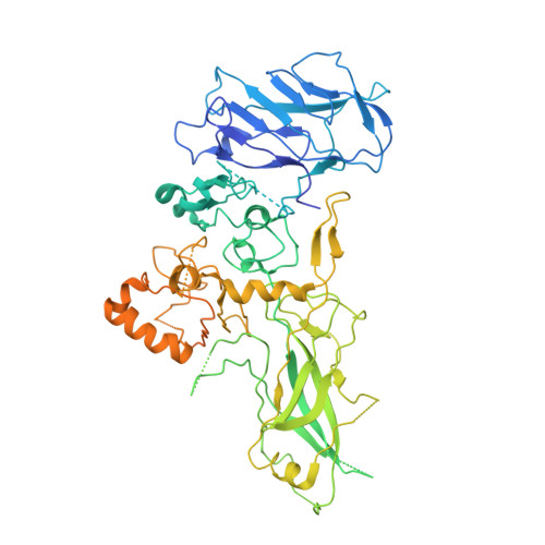
Structure and Action of the Binary C2 Toxin from Clostridium Botulinum.
Schleberger, C., Hochmann, H., Barth, H., Aktories, K., Schulz, G.E.(2006) J Mol Biol 364: 705
- PubMed: 17027031
- DOI: https://doi.org/10.1016/j.jmb.2006.09.002
- Primary Citation of Related Structures:
2J3V, 2J3X, 2J3Z, 2J42 - PubMed Abstract:
C2 toxin from Clostridium botulinum is composed of the enzyme component C2-I, which ADP-ribosylates actin, and the binding and translocation component C2-II, responsible for the interaction with eukaryotic cell receptors and the following endocytosis. Three C2-I crystal structures at resolutions of up to 1.75 A are presented together with a crystal structure of C2-II at an appreciably lower resolution and a model of the prepore formed by fragment C2-IIa. The C2-I structure was determined at pH 3.0 and at pH 6.1. The structural differences are small, indicating that C2-I does not unfold, even at a pH value as low as 3.0. The ADP-ribosyl transferase activity of C2-I was determined for alpha and beta/gamma-actin and related to that of Iota toxin and of mutant S361R of C2-I that introduced the arginine observed in Iota toxin. The substantial activity differences between alpha and beta/gamma-actin cannot be explained by the protein structures currently available. The structure of the transport component C2-II at pH 4.3 was established by molecular replacement using a model of the protective antigen of anthrax toxin at pH 6.0. The C-terminal receptor-binding domain of C2-II could not be located but was present in the crystals. It may be mobile. The relative orientation and positions of the four other domains of C2-II do not differ much from those of the protective antigen, indicating that no large conformational changes occur between pH 4.3 and pH 6.0. A model of the C2-IIa prepore structure was constructed based on the corresponding assembly of the protective antigen. It revealed a surprisingly large number of asparagine residues lining the pore. The interaction between C2-I and C2-IIa and the translocation of C2-I into the target cell are discussed.
Organizational Affiliation:
Institut für Organische Chemie und Biochemie, Albert-Ludwigs-Universität, Albertstr. 21, D-79104 Freiburg im Breisgau, Germany.