2J3M

PROLYL-TRNA SYNTHETASE FROM ENTEROCOCCUS FAECALIS COMPLEXED WITH ATP, manganese and prolinol


Experimental Data Snapshot

  • Method: X-RAY DIFFRACTION
  • Resolution: 2.30 Å
  • R-Value Free: 0.260 
  • R-Value Work: 0.196 
  • R-Value Observed: 0.198 

wwPDB Validation   3D Report Full Report


Ligand Structure Quality Assessment 


This is version 1.3 of the entry. See complete history


Literature

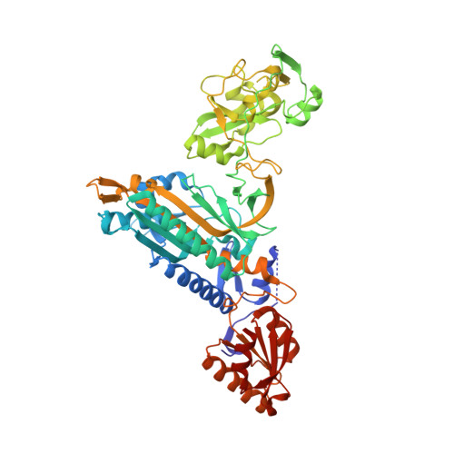
Structures of Two Bacterial Prolyl-tRNA Synthetases with and without a Cis-Editing Domain.

Crepin, T.Yaremchuk, A.Tukalo, M.Cusack, S.

(2006) Structure 14: 1511

  • DOI: https://doi.org/10.1016/j.str.2006.08.007
  • Primary Citation of Related Structures:  
    2I4L, 2I4M, 2I4N, 2I4O, 2J3L, 2J3M

  • PubMed Abstract: 

    Prolyl-tRNA synthetases (ProRSs) are unique among synthetases in that they have diverse architectures, notably the variable presence of a cis-editing domain homologous to the freestanding deacylase proteins YbaK and ProX. Here, we describe crystal structures of two bacterial ProRSs from the pathogen Enterococcus faecalis, which possesses an editing domain, and from Rhodopseudomonas palustris, which does not. We compare the overall structure and binding mode of ATP and prolyl-adenylate with those of the archael/eukaryote-type ProRS from Thermus thermophilus. Although structurally more homologous to YbaK, which preferentially hydrolyzes Cys-tRNA(Pro), the editing domain of E. faecalis ProRS possesses key elements similar to ProX, with which it shares the activity of hydrolyzing Ala-tRNA(Pro). The structures give insight into the complex evolution of ProRSs, the mechanism of editing, and structural differences between prokaryotic- and eukaryotic-type ProRSs that can be exploited for antibiotic design.


  • Organizational Affiliation

    European Molecular Biology Laboratory, Grenoble Outstation, 6 rue Jules Horowitz, BP 181, F-38042 Grenoble Cedex 9, France.


Macromolecules
Find similar proteins by:  (by identity cutoff)  |  3D Structure
Entity ID: 1
MoleculeChains Sequence LengthOrganismDetailsImage
PROLYL-TRNA SYNTHETASE
A, B
572Enterococcus faecalisMutation(s): 0 
EC: 6.1.1.15
UniProt
Find proteins for Q831W7 (Enterococcus faecalis (strain ATCC 700802 / V583))
Explore Q831W7 
Go to UniProtKB:  Q831W7
Entity Groups  
Sequence Clusters30% Identity50% Identity70% Identity90% Identity95% Identity100% Identity
UniProt GroupQ831W7
Sequence Annotations
Expand
  • Reference Sequence
Small Molecules
Ligands 3 Unique
IDChains Name / Formula / InChI Key2D Diagram3D Interactions
ATP
Query on ATP

Download Ideal Coordinates CCD File 
C [auth A],
G [auth B]
ADENOSINE-5'-TRIPHOSPHATE
C10 H16 N5 O13 P3
ZKHQWZAMYRWXGA-KQYNXXCUSA-N
PRI
Query on PRI

Download Ideal Coordinates CCD File 
D [auth A],
H [auth B]
PYRROLIDINE-2-CARBALDEHYDE
C5 H9 N O
JIDDDPVQQUHACU-YFKPBYRVSA-N
MN
Query on MN

Download Ideal Coordinates CCD File 
E [auth A]
F [auth A]
I [auth B]
J [auth B]
K [auth B]
E [auth A],
F [auth A],
I [auth B],
J [auth B],
K [auth B],
L [auth B]
MANGANESE (II) ION
Mn
WAEMQWOKJMHJLA-UHFFFAOYSA-N
Experimental Data & Validation

Experimental Data

  • Method: X-RAY DIFFRACTION
  • Resolution: 2.30 Å
  • R-Value Free: 0.260 
  • R-Value Work: 0.196 
  • R-Value Observed: 0.198 
  • Space Group: P 1 21 1
Unit Cell:
Length ( Å )Angle ( ˚ )
a = 68.583α = 90
b = 92.679β = 106.11
c = 101.223γ = 90
Software Package:
Software NamePurpose
REFMACrefinement
XDSdata reduction
XSCALEdata scaling
MOLREPphasing

Structure Validation

View Full Validation Report


Ligand Structure Quality Assessment 


Entry History 

Deposition Data

Revision History  (Full details and data files)

  • Version 1.0: 2006-10-11
    Type: Initial release
  • Version 1.1: 2011-05-08
    Changes: Version format compliance
  • Version 1.2: 2011-07-13
    Changes: Version format compliance
  • Version 1.3: 2023-12-13
    Changes: Data collection, Database references, Derived calculations, Other, Refinement description