2J0T

Crystal Structure of the Catalytic Domain of MMP-1 in Complex with the Inhibitory Domain of TIMP-1


Experimental Data Snapshot

  • Method: X-RAY DIFFRACTION
  • Resolution: 2.54 Å
  • R-Value Free: 0.275 
  • R-Value Work: 0.248 
  • R-Value Observed: 0.249 

wwPDB Validation   3D Report Full Report


This is version 1.2 of the entry. See complete history


Literature

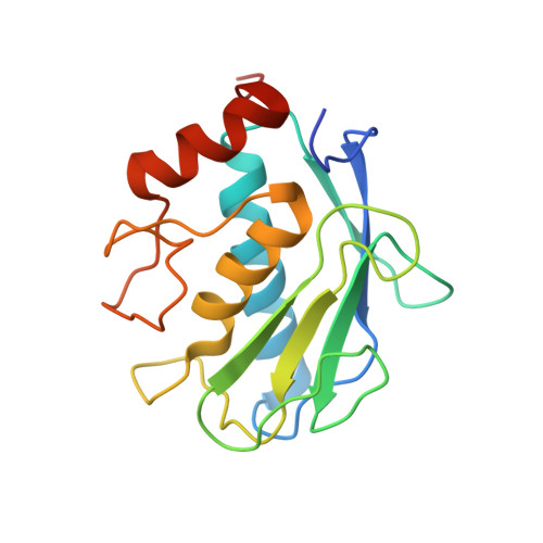
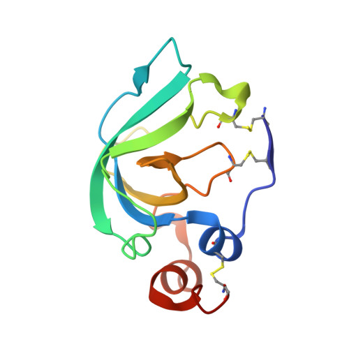
Crystal Structure of the Catalytic Domain of Matrix Metalloproteinase-1 in Complex with the Inhibitory Domain of Tissue Inhibitor of Metalloproteinase-1.

Iyer, S.Wei, S.Brew, K.Acharya, K.R.

(2007) J Biol Chem 282: 364

  • DOI: https://doi.org/10.1074/jbc.M607625200
  • Primary Citation of Related Structures:  
    2J0T

  • PubMed Abstract: 

    The mammalian collagenases are a subgroup of the matrix metalloproteinases (MMPs) that are uniquely able to cleave triple helical fibrillar collagens. Collagen breakdown is an essential part of extracellular matrix turnover in key physiological processes including morphogenesis and wound healing; however, unregulated collagenolysis is linked to important diseases such as arthritis and cancer. The tissue inhibitors of metalloproteinases (TIMPs) function in controlling the activity of MMPs, including collagenases. We report here the structure of a complex of the catalytic domain of fibroblast collagenase (MMP-1) with the N-terminal inhibitory domain of human TIMP-1 (N-TIMP-1) at 2.54 A resolution. Comparison with the previously reported structure of the TIMP-1/stromelysin-1 (MMP-3) complex shows that the mechanisms of inhibition of both MMPs are generally similar, yet there are significant differences in the protein-protein interfaces in the two complexes. Specifically, the loop between beta-strands A and B of TIMP-1 makes contact with MMP-3 but not with MMP-1, and there are marked differences in the roles of individual residues in the C-D connector of TIMP-1 in binding to the two MMPs. Structural rearrangements in the bound MMPs are also strikingly different. This is the first crystallographic structure that contains the truncated N-terminal domain of a TIMP, which shows only minor differences from the corresponding region of the full-length protein. Differences in the interactions in the two TIMP-1 complexes provide a structural explanation for the results of previous mutational studies and a basis for designing new N-TIMP-1 variants with restricted specificity.


  • Organizational Affiliation

    Department of Biology and Biochemistry, University of Bath, Claverton Down, Bath BA2 7AY, UK.


Macromolecules
Find similar proteins by:  (by identity cutoff)  |  3D Structure
Entity ID: 1
MoleculeChains Sequence LengthOrganismDetailsImage
INTERSTITIAL COLLAGENASE
A, B, C
170Homo sapiensMutation(s): 0 
EC: 3.4.24.7
UniProt & NIH Common Fund Data Resources
Find proteins for P03956 (Homo sapiens)
Explore P03956 
Go to UniProtKB:  P03956
PHAROS:  P03956
GTEx:  ENSG00000196611 
Entity Groups  
Sequence Clusters30% Identity50% Identity70% Identity90% Identity95% Identity100% Identity
UniProt GroupP03956
Sequence Annotations
Expand
  • Reference Sequence
Find similar proteins by:  (by identity cutoff)  |  3D Structure
Entity ID: 2
MoleculeChains Sequence LengthOrganismDetailsImage
METALLOPROTEINASE INHIBITOR 1
D, E, F
126Homo sapiensMutation(s): 0 
UniProt & NIH Common Fund Data Resources
Find proteins for P01033 (Homo sapiens)
Explore P01033 
Go to UniProtKB:  P01033
PHAROS:  P01033
GTEx:  ENSG00000102265 
Entity Groups  
Sequence Clusters30% Identity50% Identity70% Identity90% Identity95% Identity100% Identity
UniProt GroupP01033
Sequence Annotations
Expand
  • Reference Sequence
Small Molecules
Ligands 2 Unique
IDChains Name / Formula / InChI Key2D Diagram3D Interactions
ZN
Query on ZN

Download Ideal Coordinates CCD File 
G [auth A]
H [auth A]
L [auth B]
M [auth B]
Q [auth C]
G [auth A],
H [auth A],
L [auth B],
M [auth B],
Q [auth C],
R [auth C]
ZINC ION
Zn
PTFCDOFLOPIGGS-UHFFFAOYSA-N
CA
Query on CA

Download Ideal Coordinates CCD File 
I [auth A]
J [auth A]
K [auth A]
N [auth B]
O [auth B]
I [auth A],
J [auth A],
K [auth A],
N [auth B],
O [auth B],
P [auth B],
S [auth C],
T [auth C],
U [auth C]
CALCIUM ION
Ca
BHPQYMZQTOCNFJ-UHFFFAOYSA-N
Experimental Data & Validation

Experimental Data

  • Method: X-RAY DIFFRACTION
  • Resolution: 2.54 Å
  • R-Value Free: 0.275 
  • R-Value Work: 0.248 
  • R-Value Observed: 0.249 
  • Space Group: C 1 2 1
Unit Cell:
Length ( Å )Angle ( ˚ )
a = 158.098α = 90
b = 67.85β = 100.29
c = 86.241γ = 90
Software Package:
Software NamePurpose
REFMACrefinement
DENZOdata reduction
SCALEPACKdata scaling
AMoREphasing

Structure Validation

View Full Validation Report


Entry History 

Deposition Data

Revision History  (Full details and data files)

  • Version 1.0: 2006-10-18
    Type: Initial release
  • Version 1.1: 2011-07-13
    Changes: Advisory, Version format compliance
  • Version 1.2: 2023-12-13
    Changes: Data collection, Database references, Derived calculations, Other, Refinement description