Ab5 Subtilase Cytotoxin Inactivates the Endoplasmic Reticulum Chaperone Bip
Paton, A.W., Beddoe, T., Thorpe, C.M., Whisstock, J.C., Wilce, M.C.J., Rossjohn, J., Talbot, U.M., Paton, J.C.(2006) Nature 443: 548
- PubMed: 17024087
- DOI: https://doi.org/10.1038/nature05124
- Primary Citation of Related Structures:
2IY9 - PubMed Abstract:
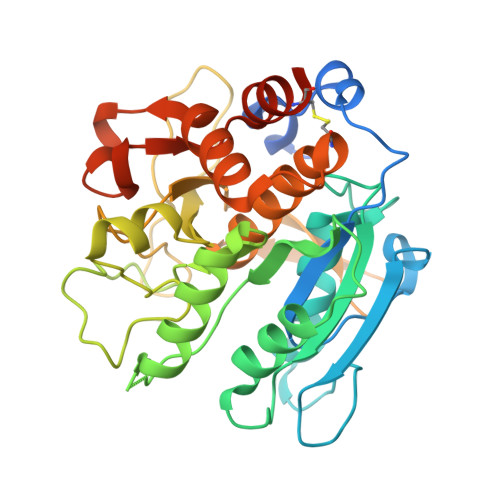
AB5 toxins are produced by pathogenic bacteria and consist of enzymatic A subunits that corrupt essential eukaryotic cell functions, and pentameric B subunits that mediate uptake into the target cell. AB5 toxins include the Shiga, cholera and pertussis toxins and a recently discovered fourth family, subtilase cytotoxin, which is produced by certain Shiga toxigenic strains of Escherichia coli. Here we show that the extreme cytotoxicity of this toxin for eukaryotic cells is due to a specific single-site cleavage of the essential endoplasmic reticulum chaperone BiP/GRP78. The A subunit is a subtilase-like serine protease; structural studies revealed an unusually deep active-site cleft, which accounts for its exquisite substrate specificity. A single amino-acid substitution in the BiP target site prevented cleavage, and co-expression of this resistant protein protected transfected cells against the toxin. BiP is a master regulator of endoplasmic reticulum function, and its cleavage by subtilase cytotoxin represents a previously unknown trigger for cell death.
Organizational Affiliation:
School of Molecular and Biomedical Science, University of Adelaide, South Australia 5005, Australia. adrienne.paton@adelaide.edu.au