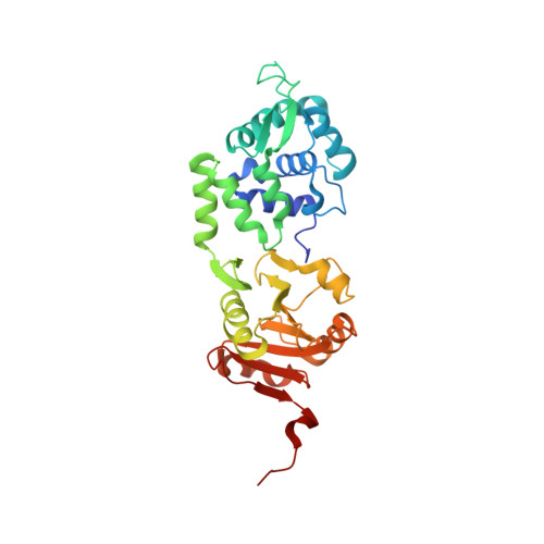
2IXS

Structure of SdaI restriction endonuclease


Experimental Data Snapshot

  • Method: X-RAY DIFFRACTION
  • Resolution: 2.00 Å
  • R-Value Free: 0.212 
  • R-Value Work: 0.178 
  • R-Value Observed: 0.182 

wwPDB Validation   3D Report Full Report


This is version 1.2 of the entry. See complete history


Literature

The Crystal Structure of the Rare-Cutting Restriction Enzyme Sdai Reveals Unexpected Domain Architecture

Tamulaitiene, G.Jakubauskas, A.Urbanke, C.Huber, R.Grazulis, S.Siksnys, V.

(2006) Structure 14: 1389

  • DOI: https://doi.org/10.1016/j.str.2006.07.002
  • Primary Citation of Related Structures:  
    2IXS

  • PubMed Abstract: 

    Rare-cutting restriction enzymes are important tools in genome analysis. We report here the crystal structure of SdaI restriction endonuclease, which is specific for the 8 bp sequence CCTGCA/GG ("/" designates the cleavage site). Unlike orthodox Type IIP enzymes, which are single domain proteins, the SdaI monomer is composed of two structural domains. The N domain contains a classical winged helix-turn-helix (wHTH) DNA binding motif, while the C domain shows a typical restriction endonuclease fold. The active site of SdaI is located within the C domain and represents a variant of the canonical PD-(D/E)XK motif. SdaI determinants of sequence specificity are clustered on the recognition helix of the wHTH motif at the N domain. The modular architecture of SdaI, wherein one domain mediates DNA binding while the other domain is predicted to catalyze hydrolysis, distinguishes SdaI from previously characterized restriction enzymes interacting with symmetric recognition sequences.


  • Organizational Affiliation

    Institute of Biotechnology, Graiciuno 8, LT-02241 Vilnius, Lithuania.


Macromolecules
Find similar proteins by:  (by identity cutoff)  |  3D Structure
Entity ID: 1
MoleculeChains Sequence LengthOrganismDetailsImage
SDAI RESTRICTION ENDONUCLEASE
A, B
323Streptomyces diastaticusMutation(s): 0 
EC: 3.1.21.4
UniProt
Find proteins for C4P954 (Streptomyces diastaticus)
Explore C4P954 
Go to UniProtKB:  C4P954
Entity Groups  
Sequence Clusters30% Identity50% Identity70% Identity90% Identity95% Identity100% Identity
UniProt GroupC4P954
Sequence Annotations
Expand
  • Reference Sequence
Small Molecules
Ligands 3 Unique
IDChains Name / Formula / InChI Key2D Diagram3D Interactions
EPE
Query on EPE

Download Ideal Coordinates CCD File 
H [auth A],
N [auth B]
4-(2-HYDROXYETHYL)-1-PIPERAZINE ETHANESULFONIC ACID
C8 H18 N2 O4 S
JKMHFZQWWAIEOD-UHFFFAOYSA-N
TRS
Query on TRS

Download Ideal Coordinates CCD File 
I [auth A],
O [auth B]
2-AMINO-2-HYDROXYMETHYL-PROPANE-1,3-DIOL
C4 H12 N O3
LENZDBCJOHFCAS-UHFFFAOYSA-O
SO4
Query on SO4

Download Ideal Coordinates CCD File 
C [auth A]
D [auth A]
E [auth A]
F [auth A]
G [auth A]
C [auth A],
D [auth A],
E [auth A],
F [auth A],
G [auth A],
J [auth B],
K [auth B],
L [auth B],
M [auth B]
SULFATE ION
O4 S
QAOWNCQODCNURD-UHFFFAOYSA-L
Experimental Data & Validation

Experimental Data

  • Method: X-RAY DIFFRACTION
  • Resolution: 2.00 Å
  • R-Value Free: 0.212 
  • R-Value Work: 0.178 
  • R-Value Observed: 0.182 
  • Space Group: C 2 2 21
Unit Cell:
Length ( Å )Angle ( ˚ )
a = 66.901α = 90
b = 237.991β = 90
c = 111.404γ = 90
Software Package:
Software NamePurpose
REFMACrefinement
MOSFLMdata reduction
SCALAdata scaling
MLPHAREphasing

Structure Validation

View Full Validation Report


Entry History 

Deposition Data

Revision History  (Full details and data files)

  • Version 1.0: 2006-09-14
    Type: Initial release
  • Version 1.1: 2011-05-08
    Changes: Version format compliance
  • Version 1.2: 2011-07-13
    Changes: Version format compliance