Atomic crystal and molecular dynamics simulation structures of human carbonic anhydrase II: insights into the proton transfer mechanism
Fisher, S.Z., Maupin, C.M., Budayova-Spano, M., Govindasamy, L., Tu, C., Agbandje-McKenna, M., Silverman, D.N., Voth, G.A., McKenna, R.(2007) Biochemistry 46: 2930-2937
- PubMed: 17319692
- DOI: https://doi.org/10.1021/bi062066y
- Primary Citation of Related Structures:
2ILI - PubMed Abstract:
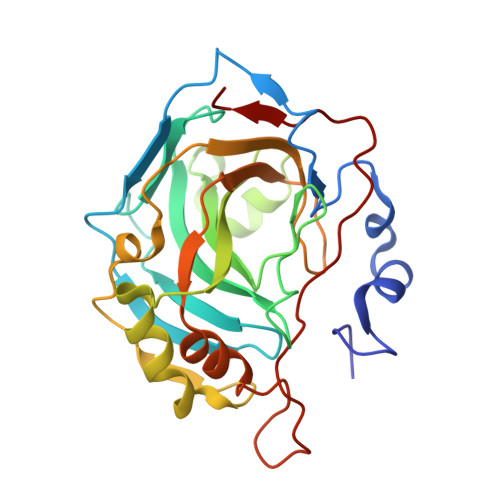
Human carbonic anhydrase II (HCA II) is a zinc-metalloenzyme that catalyzes the reversible interconversion of CO2 and HCO3-. The rate-limiting step of this catalysis is the transfer of a proton between the Zn-bound solvent molecule and residue His64. In order to fully characterize the active site structural features implicated in the proton transfer mechanism, the refined X-ray crystal structure of uncomplexed wild type HCA II to 1.05 A resolution with an Rcryst value of 12.0% and an Rfree value of 15.1% has been elucidated. This structure provides strong clues as to the pathway of the intramolecular proton transfer between the Zn-bound solvent and His64. The structure emphasizes the role of the solvent network, the unique positioning of solvent molecule W2, and the significance of the dual conformation of His64 in the active site. The structure is compared with molecular dynamics (MD) simulation calculations of the Zn-bound hydroxyl/His64+ (charged) and the Zn-bound water/His64 (uncharged) HCA II states. A comparison of the crystallographic anisotropic atomic thermal parameters and MD simulation root-mean-square fluctuation values show excellent agreement in the atomic motion observed between the two methods. It is also interesting that the observed active site solvent positions in the crystal structure are also the most probable positions of the solvent during the MD simulations. On the basis of the comparative study of the MD simulation results, the HCA II crystal structure observed is most likely in the Zn-bound water/His64 state. This conclusion is based on the following observations: His64 is mainly (80%) orientated in an inward conformation; electron density omit maps infer that His64 is not charged in an either inward or outward conformation; and the Zn-bound solvent is most likely a water molecule.
Organizational Affiliation:
Department of Biochemistry and Molecular Biology, College of Medicine, University of Florida, Gainesville, Florida 32610, USA.