2I66

Structural Basis for the Mechanistic Understanding Human CD38 Controlled Multiple Catalysis


Experimental Data Snapshot

  • Method: X-RAY DIFFRACTION
  • Resolution: 1.70 Å
  • R-Value Free: 0.229 
  • R-Value Work: 0.182 
  • R-Value Observed: 0.185 

wwPDB Validation   3D Report Full Report


This is version 1.4 of the entry. See complete history


Literature

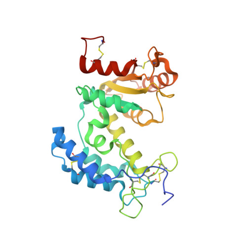
Structural basis for the mechanistic understanding of human CD38-controlled multiple catalysis.

Liu, Q.Kriksunov, I.A.Graeff, R.Munshi, C.Lee, H.C.Hao, Q.

(2006) J Biol Chem 281: 32861-32869

  • DOI: https://doi.org/10.1074/jbc.M606365200
  • Primary Citation of Related Structures:  
    2I65, 2I66, 2I67

  • PubMed Abstract: 

    The enzymatic cleavage of the nicotinamide-glycosidic bond on nicotinamide adenine dinucleotide (NAD(+)) has been proposed to go through an oxocarbenium ion-like transition state. Because of the instability of the ionic intermediate, there has been no structural report on such a transient reactive species. Human CD38 is an ectoenzyme that can use NAD(+) to synthesize two calcium-mobilizing molecules. By using NAD(+) and a surrogate substrate, NGD(+), we captured and determined crystal structures of the enzyme complexed with an intermediate, a substrate, and a product along the reaction pathway. Our results showed that the intermediate is stabilized by polar interactions with the catalytic residue Glu(226) rather than by a covalent linkage. The polar interactions between Glu(226) and the substrate 2',3'-OH groups are essential for initiating catalysis. Ser(193) was demonstrated to have a regulative role during catalysis and is likely to be involved in intermediate stabilization. In addition, a product inhibition effect by ADP-ribose (through the reorientation of the product) or GDP-ribose (through the formation of a covalently linked GDP-ribose dimer) was observed. These structural data provide insights into the understanding of multiple catalysis and clues for drug design.


  • Organizational Affiliation

    Macromolecular Diffraction Facility at the Cornell High Energy Synchrotron Source (MacCHESS), Cornell University, Ithaca, NY 14853, USA.


Macromolecules
Find similar proteins by:  (by identity cutoff)  |  3D Structure
Entity ID: 1
MoleculeChains Sequence LengthOrganismDetailsImage
ADP-ribosyl cyclase 1
A, B
262Homo sapiensMutation(s): 5 
Gene Names: CD38
EC: 3.2.2.5
UniProt & NIH Common Fund Data Resources
Find proteins for P28907 (Homo sapiens)
Explore P28907 
Go to UniProtKB:  P28907
PHAROS:  P28907
GTEx:  ENSG00000004468 
Entity Groups  
Sequence Clusters30% Identity50% Identity70% Identity90% Identity95% Identity100% Identity
UniProt GroupP28907
Sequence Annotations
Expand
  • Reference Sequence
Small Molecules
Ligands 2 Unique
IDChains Name / Formula / InChI Key2D Diagram3D Interactions
G1R
Query on G1R

Download Ideal Coordinates CCD File 
C [auth A],
D [auth A],
E [auth B]
[(2R,3R,4R,5R)-5-(2-AMINO-6-OXO-1,6-DIHYDRO-9H-PURIN-9-YL)-3,4-DIHYDROXYTETRAHYDROFURAN-2-YL]METHYL [(2R,3S,4R,5S)-3,4,5-TRIHYDROXYTETRAHYDROFURAN-2-YL]METHYL DIHYDROGEN DIPHOSPHATE
C15 H23 N5 O15 P2
JSQOXYZOPSTQIZ-NQRHNBEYSA-N
G2R
Query on G2R

Download Ideal Coordinates CCD File 
F [auth B][(2R,3R,4R,5R)-5-(2-AMINO-6-OXO-1,6-DIHYDRO-9H-PURIN-9-YL)-3,4-DIHYDROXYTETRAHYDROFURAN-2-YL]METHYL [(2R,3S,4S)-3,4-DIHYDROXYTETRAHYDROFURAN-2-YL]METHYL DIHYDROGEN DIPHOSPHATE
C15 H23 N5 O14 P2
NOLLEWZDVQOFJY-DCLLCRAESA-N
Experimental Data & Validation

Experimental Data

  • Method: X-RAY DIFFRACTION
  • Resolution: 1.70 Å
  • R-Value Free: 0.229 
  • R-Value Work: 0.182 
  • R-Value Observed: 0.185 
  • Space Group: P 1
Unit Cell:
Length ( Å )Angle ( ˚ )
a = 41.71α = 105.93
b = 52.812β = 91.8
c = 65.424γ = 95.22
Software Package:
Software NamePurpose
REFMACrefinement
ADSCdata collection
HKL-2000data reduction
HKL-2000data scaling
MOLREPphasing

Structure Validation

View Full Validation Report


Entry History 

Deposition Data

Revision History  (Full details and data files)

  • Version 1.0: 2006-09-05
    Type: Initial release
  • Version 1.1: 2008-05-01
    Changes: Version format compliance
  • Version 1.2: 2011-07-13
    Changes: Version format compliance
  • Version 1.3: 2021-10-20
    Changes: Database references, Derived calculations
  • Version 1.4: 2023-08-30
    Changes: Data collection, Refinement description