2I5F

Crystal structure of the C-terminal PH domain of pleckstrin in complex with D-myo-Ins(1,2,3,5,6)P5


Experimental Data Snapshot

  • Method: X-RAY DIFFRACTION
  • Resolution: 1.35 Å
  • R-Value Free: 0.208 
  • R-Value Work: 0.179 
  • R-Value Observed: 0.179 

wwPDB Validation   3D Report Full Report


This is version 1.2 of the entry. See complete history


Literature

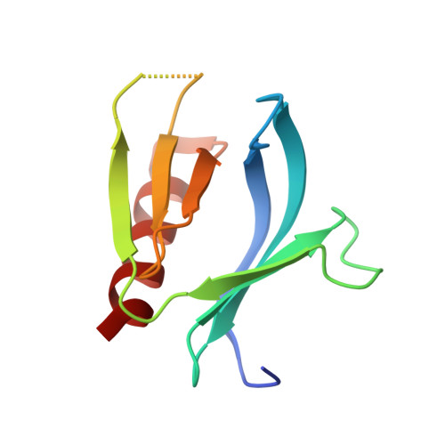
Structural analysis of the carboxy terminal PH domain of pleckstrin bound to D-myo-inositol 1,2,3,5,6-pentakisphosphate.

Jackson, S.G.Zhang, Y.Haslam, R.J.Junop, M.S.

(2007) BMC Struct Biol 7: 80-80

  • DOI: https://doi.org/10.1186/1472-6807-7-80
  • Primary Citation of Related Structures:  
    2I5C, 2I5F

  • PubMed Abstract: 

    Pleckstrin homology (PH) domains are one of the most prevalent domains in the human proteome and represent the major phosphoinositide-binding module. These domains are often found in signaling proteins and function predominately by targeting their host proteins to the cell membrane. Inositol phosphates, which are structurally similar to phosphoinositides, are not only known to play a role as signaling molecules but are also capable of being bound by PH domains.


  • Organizational Affiliation

    Department of Biochemistry and Biomedical Sciences, McMaster University, Hamilton, Canada. jacksosg@mcmaster.ca


Macromolecules
Find similar proteins by:  (by identity cutoff)  |  3D Structure
Entity ID: 1
MoleculeChains Sequence LengthOrganismDetailsImage
Pleckstrin109Homo sapiensMutation(s): 0 
Gene Names: PLEKP47
UniProt & NIH Common Fund Data Resources
Find proteins for P08567 (Homo sapiens)
Explore P08567 
Go to UniProtKB:  P08567
PHAROS:  P08567
GTEx:  ENSG00000115956 
Entity Groups  
Sequence Clusters30% Identity50% Identity70% Identity90% Identity95% Identity100% Identity
UniProt GroupP08567
Sequence Annotations
Expand
  • Reference Sequence
Small Molecules
Ligands 1 Unique
IDChains Name / Formula / InChI Key2D Diagram3D Interactions
5IP
Query on 5IP

Download Ideal Coordinates CCD File 
B [auth A](1R,2R,3R,4R,5S,6S)-6-HYDROXYCYCLOHEXANE-1,2,3,4,5-PENTAYL PENTAKIS[DIHYDROGEN (PHOSPHATE)]
C6 H17 O21 P5
CTPQAXVNYGZUAJ-RTWKJESVSA-N
Binding Affinity Annotations 
IDSourceBinding Affinity
5IP PDBBind:  2I5F IC50: 7500 (nM) from 1 assay(s)
Binding MOAD:  2I5F IC50: 2200 (nM) from 1 assay(s)
Experimental Data & Validation

Experimental Data

  • Method: X-RAY DIFFRACTION
  • Resolution: 1.35 Å
  • R-Value Free: 0.208 
  • R-Value Work: 0.179 
  • R-Value Observed: 0.179 
  • Space Group: P 21 21 21
Unit Cell:
Length ( Å )Angle ( ˚ )
a = 32.15α = 90
b = 47.7β = 90
c = 64.06γ = 90
Software Package:
Software NamePurpose
REFMACrefinement
CBASSdata collection
d*TREKdata reduction
d*TREKdata scaling
MOLREPphasing

Structure Validation

View Full Validation Report


Entry History 

Deposition Data

Revision History  (Full details and data files)

  • Version 1.0: 2007-08-07
    Type: Initial release
  • Version 1.1: 2011-07-13
    Changes: Version format compliance
  • Version 1.2: 2024-02-21
    Changes: Data collection, Database references, Derived calculations