


The crystal structure of the class C acid phosphatase from Bacillus anthracis
Felts, R.L., Tanner, J.J.To be published.
Experimental Data Snapshot
wwPDB Validation 3D Report Full Report

Entity ID: 1 | |||||
|---|---|---|---|---|---|
| Molecule | Chains | Sequence Length | Organism | Details | Image |
| acid phosphatase | 258 | Bacillus anthracis | Mutation(s): 0 EC: 3.1.3.2 | 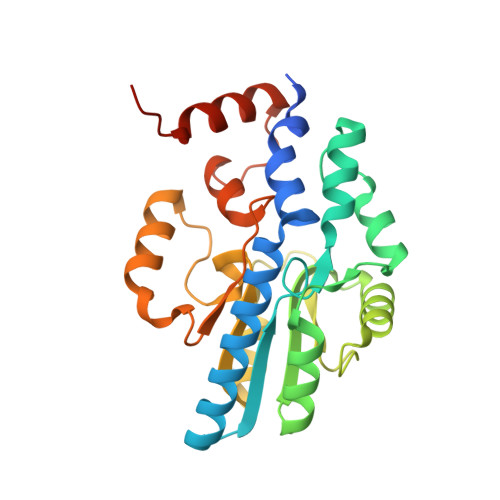 | |
UniProt | |||||
Find proteins for A0A6L7HE29 (Bacillus anthracis) Explore A0A6L7HE29 Go to UniProtKB: A0A6L7HE29 | |||||
Entity Groups | |||||
| Sequence Clusters | 30% Identity50% Identity70% Identity90% Identity95% Identity100% Identity | ||||
| UniProt Group | A0A6L7HE29 | ||||
Sequence AnnotationsExpand | |||||
| |||||
| Ligands 2 Unique | |||||
|---|---|---|---|---|---|
| ID | Chains | Name / Formula / InChI Key | 2D Diagram | 3D Interactions | |
| WO4 Query on WO4 | D [auth A], F [auth B] | TUNGSTATE(VI)ION O4 W PBYZMCDFOULPGH-UHFFFAOYSA-N |  | ||
| MG Query on MG | C [auth A], E [auth B] | MAGNESIUM ION Mg JLVVSXFLKOJNIY-UHFFFAOYSA-N |  | ||
| Length ( Å ) | Angle ( ˚ ) |
|---|---|
| a = 53 | α = 90 |
| b = 89.94 | β = 90 |
| c = 104.12 | γ = 90 |
| Software Name | Purpose |
|---|---|
| d*TREK | data scaling |
| SOLVE | phasing |
| RESOLVE | phasing |
| REFMAC | refinement |
| PDB_EXTRACT | data extraction |
| d*TREK | data reduction |
RCSB PDB (citation) is hosted by
RCSB PDB is a member of the

