


High resolution crystal structures of RafE from Streptococcus pneumoniae
Paterson, N.G., Riboldi-Tunnicliffe, A., Mitchell, T.J., Isaacs, N.W.To be published.
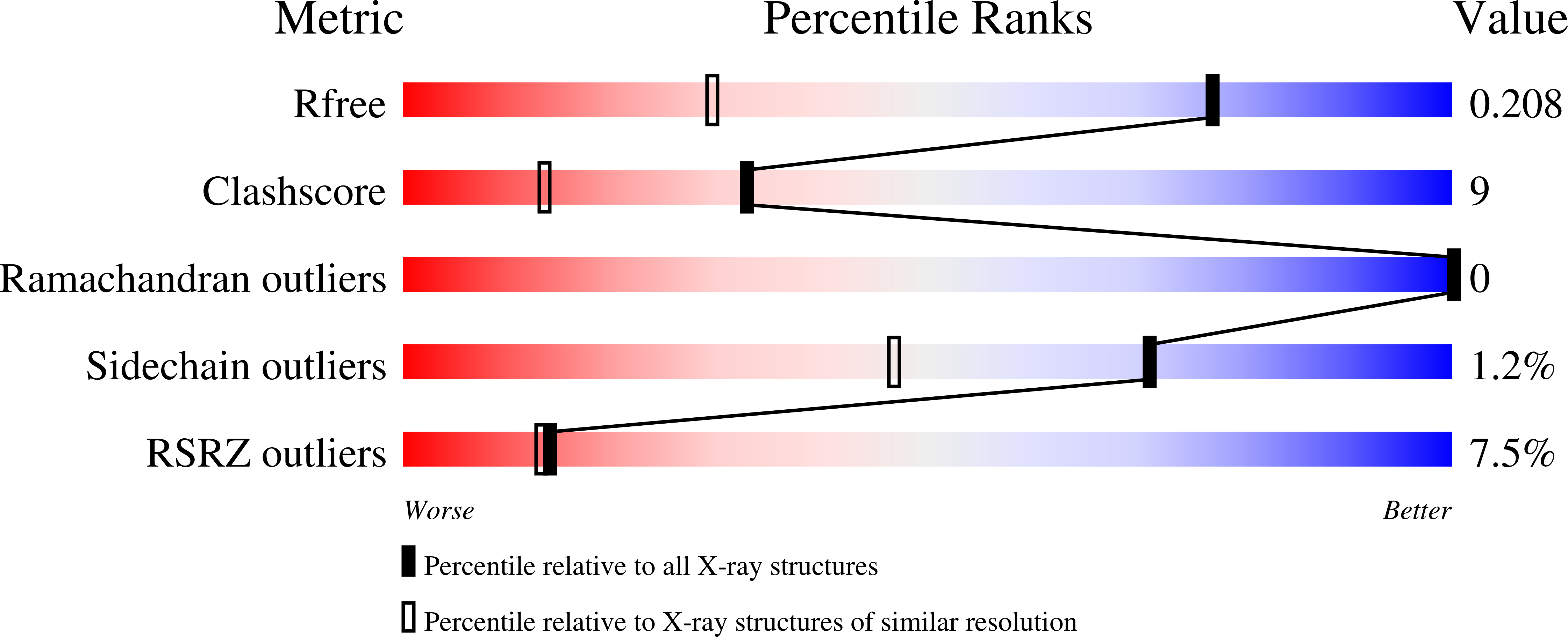
Experimental Data Snapshot
wwPDB Validation 3D Report Full Report
Entity ID: 1 | |||||
|---|---|---|---|---|---|
| Molecule | Chains | Sequence Length | Organism | Details | Image |
| Sugar ABC transporter, sugar-binding protein | 388 | Streptococcus pneumoniae TIGR4 | Mutation(s): 0 Gene Names: rafE | 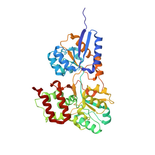 | |
UniProt | |||||
Find proteins for A0A0H2URJ7 (Streptococcus pneumoniae serotype 4 (strain ATCC BAA-334 / TIGR4)) Explore A0A0H2URJ7 Go to UniProtKB: A0A0H2URJ7 | |||||
Entity Groups | |||||
| Sequence Clusters | 30% Identity50% Identity70% Identity90% Identity95% Identity100% Identity | ||||
| UniProt Group | A0A0H2URJ7 | ||||
Sequence AnnotationsExpand | |||||
| |||||
| Ligands 2 Unique | |||||
|---|---|---|---|---|---|
| ID | Chains | Name / Formula / InChI Key | 2D Diagram | 3D Interactions | |
| ACT Query on ACT | C [auth A], D [auth A], E [auth A] | ACETATE ION C2 H3 O2 QTBSBXVTEAMEQO-UHFFFAOYSA-M |  | ||
| NA Query on NA | B [auth A] | SODIUM ION Na FKNQFGJONOIPTF-UHFFFAOYSA-N |  | ||
| Length ( Å ) | Angle ( ˚ ) |
|---|---|
| a = 73.754 | α = 90 |
| b = 77.7 | β = 90 |
| c = 79.226 | γ = 90 |
| Software Name | Purpose |
|---|---|
| d*TREK | data scaling |
| PHASER | phasing |
| REFMAC | refinement |
| PDB_EXTRACT | data extraction |
| d*TREK | data reduction |
RCSB PDB (citation) is hosted by
RCSB PDB is a member of the

