2HNT

CRYSTALLOGRAPHIC STRUCTURE OF HUMAN GAMMA-THROMBIN


Experimental Data Snapshot

  • Method: X-RAY DIFFRACTION
  • Resolution: 2.50 Å
  • R-Value Observed: 0.156 

wwPDB Validation   3D Report Full Report


This is version 1.2 of the entry. See complete history


Literature

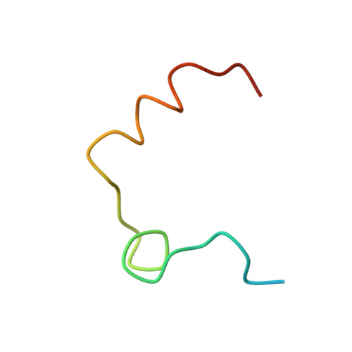
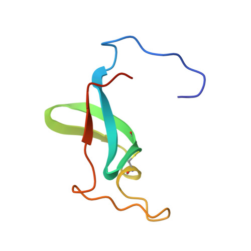
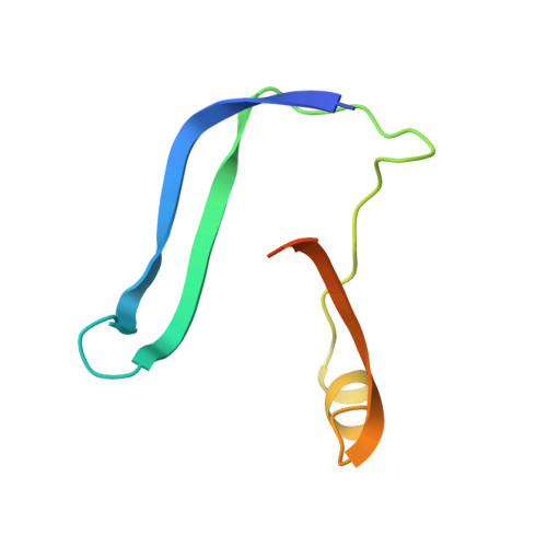
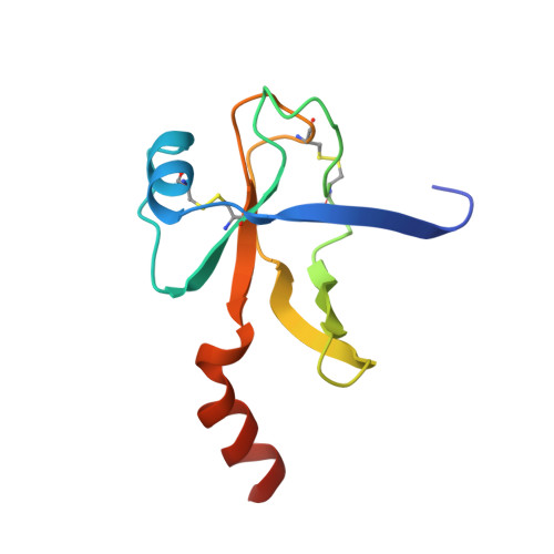
Crystallographic structure of human gamma-thrombin.

Rydel, T.J.Yin, M.Padmanabhan, K.P.Blankenship, D.T.Cardin, A.D.Correa, P.E.Fenton 2nd., J.W.Tulinsky, A.

(1994) J Biol Chem 269: 22000-22006

  • Primary Citation of Related Structures:  
    2HNT

  • PubMed Abstract: 

    In an effort to prepare crystals and determine the structure of alpha-thrombin complexed to a synthetic peptide inhibitor (MDL-28050) of the hirudin 54-65 COOH-terminal region, it was discovered that the crystals were not those of the complex but of gamma-thrombin. Gel electrophoresis studies revealed that autolytic degradation had occurred prior to crystallization. NH2-terminal sequence analysis of these autolytic fragments confirmed the gamma-thrombin product (cleavages at Arg75-Tyr76 and/or Arg77A-Asn78, and Lys149E-Gly150; chymotrypsinogen numbering) with a minor amount of another autolysis product, beta-thrombin (first two cleavages only). The final structure has an R-factor of 0.156 for 7.0-2.5-A data, and includes 186 water molecules. A comparison of gamma-thrombin with the thrombin structure in the alpha-thrombin-hirugen complex revealed that the two structures agreed well (r.m.s. delta = 0.39 A for main chain atoms). These structures possess uninhibited active sites where the disposition of the catalytic triad residues is nearly identical. The electron density in the vicinity of the gamma-thrombin cleavage regions is poor, and only becomes well-defined several residues prior to and after the actual cleavage sites. The extensive disorder evoked by beta-cleavage(s) in the Lys70-Glu80 loop region indicates that this part of the molecule is severely disrupted by autolysis and is the reason exosite functions are dramatically impaired in beta-and gamma-thrombin. Since autolysis did not lead to a major reorganization of the folded structure of alpha-thrombin, the likely structural features of the interaction of thrombin substrate with thrombin enzyme during beta-cleavage have been modeled by docking the exosite region of one molecule at the active site of another.


  • Organizational Affiliation

    Department of Chemistry, Michigan State University, East Lansing 48824-1322.


Macromolecules
Find similar proteins by:  (by identity cutoff)  |  3D Structure
Entity ID: 1
MoleculeChains Sequence LengthOrganismDetailsImage
GAMMA-THROMBINA [auth L]36Homo sapiensMutation(s): 0 
UniProt & NIH Common Fund Data Resources
Find proteins for P00734 (Homo sapiens)
Explore P00734 
Go to UniProtKB:  P00734
PHAROS:  P00734
GTEx:  ENSG00000180210 
Entity Groups  
Sequence Clusters30% Identity50% Identity70% Identity90% Identity95% Identity100% Identity
UniProt GroupP00734
Sequence Annotations
Expand
  • Reference Sequence
Find similar proteins by:  (by identity cutoff)  |  3D Structure
Entity ID: 2
MoleculeChains Sequence LengthOrganismDetailsImage
GAMMA-THROMBINB [auth C]70Homo sapiensMutation(s): 0 
UniProt & NIH Common Fund Data Resources
Find proteins for P00734 (Homo sapiens)
Explore P00734 
Go to UniProtKB:  P00734
PHAROS:  P00734
GTEx:  ENSG00000180210 
Entity Groups  
Sequence Clusters30% Identity50% Identity70% Identity90% Identity95% Identity100% Identity
UniProt GroupP00734
Sequence Annotations
Expand
  • Reference Sequence
Find similar proteins by:  (by identity cutoff)  |  3D Structure
Entity ID: 3
MoleculeChains Sequence LengthOrganismDetailsImage
GAMMA-THROMBINC [auth E]81Homo sapiensMutation(s): 0 
UniProt & NIH Common Fund Data Resources
Find proteins for P00734 (Homo sapiens)
Explore P00734 
Go to UniProtKB:  P00734
PHAROS:  P00734
GTEx:  ENSG00000180210 
Entity Groups  
Sequence Clusters30% Identity50% Identity70% Identity90% Identity95% Identity100% Identity
UniProt GroupP00734
Sequence Annotations
Expand
  • Reference Sequence
Find similar proteins by:  (by identity cutoff)  |  3D Structure
Entity ID: 4
MoleculeChains Sequence LengthOrganismDetailsImage
GAMMA-THROMBIND [auth F]105Homo sapiensMutation(s): 0 
UniProt & NIH Common Fund Data Resources
Find proteins for P00734 (Homo sapiens)
Explore P00734 
Go to UniProtKB:  P00734
PHAROS:  P00734
GTEx:  ENSG00000180210 
Entity Groups  
Sequence Clusters30% Identity50% Identity70% Identity90% Identity95% Identity100% Identity
UniProt GroupP00734
Sequence Annotations
Expand
  • Reference Sequence
Experimental Data & Validation

Experimental Data

  • Method: X-RAY DIFFRACTION
  • Resolution: 2.50 Å
  • R-Value Observed: 0.156 
  • Space Group: C 1 2 1
Unit Cell:
Length ( Å )Angle ( ˚ )
a = 126.45α = 90
b = 48.23β = 96.2
c = 52.43γ = 90
Software Package:
Software NamePurpose
PROFFTrefinement

Structure Validation

View Full Validation Report


Entry History 

Deposition Data

  • Released Date: 1994-11-30 
  • Deposition Author(s): Tulinsky, A.

Revision History  (Full details and data files)

  • Version 1.0: 1994-11-30
    Type: Initial release
  • Version 1.1: 2008-03-03
    Changes: Version format compliance
  • Version 1.2: 2011-07-13
    Changes: Version format compliance