2GTG

Crystal Structure of Human Saposin C


Experimental Data Snapshot

  • Method: X-RAY DIFFRACTION
  • Resolution: 2.40 Å
  • R-Value Free: 0.283 
  • R-Value Work: 0.211 
  • R-Value Observed: 0.221 

wwPDB Validation   3D Report Full Report


This is version 1.3 of the entry. See complete history


Literature

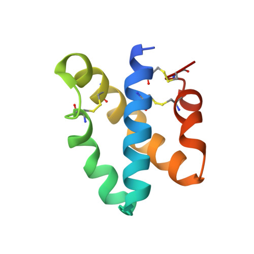
Crystal structures of saposins A and C.

Ahn, V.E.Leyko, P.Alattia, J.R.Chen, L.Prive, G.G.

(2006) Protein Sci 15: 1849-1857

  • DOI: https://doi.org/10.1110/ps.062256606
  • Primary Citation of Related Structures:  
    2DOB, 2GTG

  • PubMed Abstract: 

    Saposins A and C are sphingolipid activator proteins required for the lysosomal breakdown of galactosylceramide and glucosylceramide, respectively. The saposins interact with lipids, leading to an enhanced accessibility of the lipid headgroups to their cognate hydrolases. We have determined the crystal structures of human saposins A and C to 2.0 Angstroms and 2.4 Angstroms, respectively, and both reveal the compact, monomeric saposin fold. We confirmed that these two proteins were monomeric in solution at pH 7.0 by analytical centrifugation. However, at pH 4.8, in the presence of the detergent C(8)E(5), saposin A assembled into dimers, while saposin C formed trimers. Saposin B was dimeric under all conditions tested. The self-association of the saposins is likely to be relevant to how these small proteins interact with lipids, membranes, and hydrolase enzymes.


  • Organizational Affiliation

    Department of Medical Biophysics, University of Toronto, Canada.


Macromolecules
Find similar proteins by:  (by identity cutoff)  |  3D Structure
Entity ID: 1
MoleculeChains Sequence LengthOrganismDetailsImage
Proactivator polypeptide83Homo sapiensMutation(s): 0 
Gene Names: PSAP
UniProt & NIH Common Fund Data Resources
Find proteins for P07602 (Homo sapiens)
Explore P07602 
Go to UniProtKB:  P07602
PHAROS:  P07602
GTEx:  ENSG00000197746 
Entity Groups  
Sequence Clusters30% Identity50% Identity70% Identity90% Identity95% Identity100% Identity
UniProt GroupP07602
Sequence Annotations
Expand
  • Reference Sequence
Experimental Data & Validation

Experimental Data

  • Method: X-RAY DIFFRACTION
  • Resolution: 2.40 Å
  • R-Value Free: 0.283 
  • R-Value Work: 0.211 
  • R-Value Observed: 0.221 
  • Space Group: P 63
Unit Cell:
Length ( Å )Angle ( ˚ )
a = 53.204α = 90
b = 53.204β = 90
c = 52.46γ = 120
Software Package:
Software NamePurpose
REFMACrefinement
DENZOdata reduction
SCALEPACKdata scaling
AMoREphasing

Structure Validation

View Full Validation Report


Entry History 

Deposition Data

Revision History  (Full details and data files)

  • Version 1.0: 2006-07-25
    Type: Initial release
  • Version 1.1: 2008-05-01
    Changes: Version format compliance
  • Version 1.2: 2011-07-13
    Changes: Advisory, Version format compliance
  • Version 1.3: 2023-08-30
    Changes: Data collection, Database references, Refinement description