2GP9

Crystal structure of the slow form of thrombin in a self-inhibited conformation


Experimental Data Snapshot

  • Method: X-RAY DIFFRACTION
  • Resolution: 1.87 Å
  • R-Value Free: 0.218 
  • R-Value Work: 0.195 

wwPDB Validation   3D Report Full Report


This is version 1.4 of the entry. See complete history


Literature

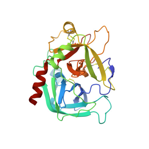
Crystal structure of thrombin in a self-inhibited conformation.

Pineda, A.O.Chen, Z.W.Bah, A.Garvey, L.C.Mathews, F.S.Di Cera, E.

(2006) J Biol Chem 281: 32922-32928

  • DOI: https://doi.org/10.1074/jbc.M605530200
  • Primary Citation of Related Structures:  
    2GP9

  • PubMed Abstract: 

    The activating effect of Na(+) on thrombin is allosteric and depends on the conformational transition from a low activity Na(+)-free (slow) form to a high activity Na(+)-bound (fast) form. The structures of these active forms have been solved. Recent structures of thrombin obtained in the absence of Na(+) have also documented inactive conformations that presumably exist in equilibrium with the active slow form. The validity of these inactive slow form structures, however, is called into question by the presence of packing interactions involving the Na(+) site and the active site regions. Here, we report a 1.87A resolution structure of thrombin in the absence of inhibitors and salts with a single molecule in the asymmetric unit and devoid of significant packing interactions in regions involved in the allosteric slow --> fast transition. The structure shows an unprecedented self-inhibited conformation where Trp-215 and Arg-221a relocate >10A to occlude the active site and the primary specificity pocket, and the guanidinium group of Arg-187 penetrates the protein core to fill the empty Na(+)-binding site. The extreme mobility of Trp-215 was investigated further with the W215P mutation. Remarkably, the mutation significantly compromises cleavage of the anticoagulant protein C but has no effect on the hydrolysis of fibrinogen and PAR1. These findings demonstrate that thrombin may assume an inactive conformation in the absence of Na(+) and that its procoagulant and anticoagulant activities are closely linked to the mobility of residue 215.


  • Organizational Affiliation

    Department of Biochemistry and Molecular Biophysics, Washington University School of Medicine, St. Louis, MO 63110, USA.


Macromolecules
Find similar proteins by:  (by identity cutoff)  |  3D Structure
Entity ID: 1
MoleculeChains Sequence LengthOrganismDetailsImage
Prothrombin36Homo sapiensMutation(s): 0 
Gene Names: F2
EC: 3.4.21.5
UniProt & NIH Common Fund Data Resources
Find proteins for P00734 (Homo sapiens)
Explore P00734 
Go to UniProtKB:  P00734
PHAROS:  P00734
GTEx:  ENSG00000180210 
Entity Groups  
Sequence Clusters30% Identity50% Identity70% Identity90% Identity95% Identity100% Identity
UniProt GroupP00734
Sequence Annotations
Expand
  • Reference Sequence
Find similar proteins by:  (by identity cutoff)  |  3D Structure
Entity ID: 2
MoleculeChains Sequence LengthOrganismDetailsImage
Prothrombin259Homo sapiensMutation(s): 1 
Gene Names: F2
EC: 3.4.21.5
UniProt & NIH Common Fund Data Resources
Find proteins for P00734 (Homo sapiens)
Explore P00734 
Go to UniProtKB:  P00734
PHAROS:  P00734
GTEx:  ENSG00000180210 
Entity Groups  
Sequence Clusters30% Identity50% Identity70% Identity90% Identity95% Identity100% Identity
UniProt GroupP00734
Sequence Annotations
Expand
  • Reference Sequence
Small Molecules
Ligands 1 Unique
IDChains Name / Formula / InChI Key2D Diagram3D Interactions
EPE
Query on EPE

Download Ideal Coordinates CCD File 
C [auth B]4-(2-HYDROXYETHYL)-1-PIPERAZINE ETHANESULFONIC ACID
C8 H18 N2 O4 S
JKMHFZQWWAIEOD-UHFFFAOYSA-N
Experimental Data & Validation

Experimental Data

  • Method: X-RAY DIFFRACTION
  • Resolution: 1.87 Å
  • R-Value Free: 0.218 
  • R-Value Work: 0.195 
  • Space Group: P 43
Unit Cell:
Length ( Å )Angle ( ˚ )
a = 57.892α = 90
b = 57.892β = 90
c = 119.937γ = 90
Software Package:
Software NamePurpose
HKL-2000data collection
SCALEPACKdata scaling
MOLREPphasing
CNSrefinement
HKL-2000data reduction

Structure Validation

View Full Validation Report


Entry History 

Deposition Data

Revision History  (Full details and data files)

  • Version 1.0: 2006-09-12
    Type: Initial release
  • Version 1.1: 2008-05-01
    Changes: Version format compliance
  • Version 1.2: 2011-07-13
    Changes: Version format compliance
  • Version 1.3: 2021-10-20
    Changes: Database references, Derived calculations
  • Version 1.4: 2023-08-30
    Changes: Data collection, Refinement description