2GP8

NMR SOLUTION STRUCTURE OF THE COAT PROTEIN-BINDING DOMAIN OF BACTERIOPHAGE P22 SCAFFOLDING PROTEIN


Experimental Data Snapshot

  • Method: SOLUTION NMR
  • Conformers Calculated: 100 
  • Conformers Submitted: 
  • Selection Criteria: LEAST RESTRAINT VIOLATION 

wwPDB Validation   3D Report Full Report

Currently 2GP8 does not have a validation slider image.


This is version 1.4 of the entry. See complete history


Literature

Structure of the coat protein-binding domain of the scaffolding protein from a double-stranded DNA virus.

Sun, Y.Parker, M.H.Weigele, P.Casjens, S.Prevelige Jr., P.E.Krishna, N.R.

(2000) J Mol Biol 297: 1195-1202

  • DOI: https://doi.org/10.1006/jmbi.2000.3620
  • Primary Citation of Related Structures:  
    1GP8, 2GP8

  • PubMed Abstract: 

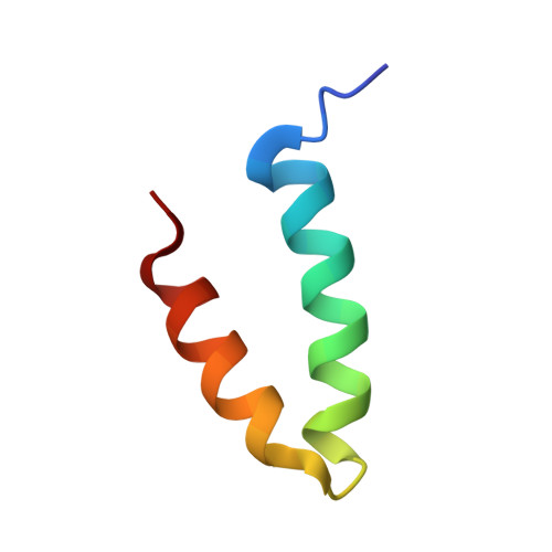
    Scaffolding proteins are required for high fidelity assembly of most high T number dsDNA viruses such as the large bacteriophages, and the herpesvirus family. They function by transiently binding and positioning the coat protein subunits during capsid assembly. In both bacteriophage P22 and the herpesviruses the extreme scaffold C terminus is highly charged, is predicted to be an amphipathic alpha-helix, and is sufficient to bind the coat protein, suggesting a common mode of action. NMR studies show that the coat protein-binding domain of P22 scaffolding protein exhibits a helix-loop-helix motif stabilized by a hydrophobic core. One face of the motif is characterized by a high density of positive charges that could interact with the coat protein through electrostatic interactions. Results from previous studies with a truncation fragment and the observed salt sensitivity of the assembly process are explained by the NMR structure.


  • Organizational Affiliation

    Comprehensive Cancer Center, Birmingham, AL, 35294, USA.


Macromolecules
Find similar proteins by:  (by identity cutoff)  |  3D Structure
Entity ID: 1
MoleculeChains Sequence LengthOrganismDetailsImage
PROTEIN (SCAFFOLDING PROTEIN)40Lederbergvirus P22Mutation(s): 0 
UniProt
Find proteins for P26748 (Salmonella phage P22)
Explore P26748 
Go to UniProtKB:  P26748
Entity Groups  
Sequence Clusters30% Identity50% Identity70% Identity90% Identity95% Identity100% Identity
UniProt GroupP26748
Sequence Annotations
Expand
  • Reference Sequence
Experimental Data & Validation

Experimental Data

  • Method: SOLUTION NMR
  • Conformers Calculated: 100 
  • Conformers Submitted: 
  • Selection Criteria: LEAST RESTRAINT VIOLATION 

Structure Validation

View Full Validation Report

Currently 2GP8 does not have a validation slider image.


Entry History 

Deposition Data

Revision History  (Full details and data files)

  • Version 1.0: 1999-05-17
    Type: Initial release
  • Version 1.1: 2008-04-26
    Changes: Version format compliance
  • Version 1.2: 2011-07-13
    Changes: Version format compliance
  • Version 1.3: 2022-03-09
    Changes: Database references, Derived calculations
  • Version 1.4: 2023-12-27
    Changes: Data collection