2GBL

Crystal Structure of Full Length Circadian Clock Protein KaiC with Phosphorylation Sites


Experimental Data Snapshot

  • Method: X-RAY DIFFRACTION
  • Resolution: 2.80 Å
  • R-Value Free: 0.290 
  • R-Value Work: 0.230 
  • R-Value Observed: 0.230 

wwPDB Validation   3D Report Full Report


Ligand Structure Quality Assessment 


This is version 1.3 of the entry. See complete history


Literature

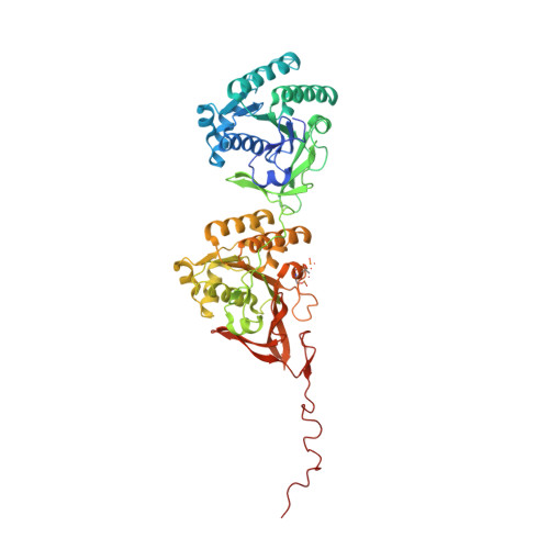
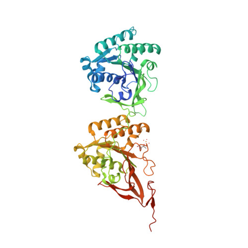
Analysis of KaiA-KaiC protein interactions in the cyano-bacterial circadian clock using hybrid structural methods.

Pattanayek, R.Williams, D.R.Pattanayek, S.Xu, Y.Mori, T.Johnson, C.H.Stewart, P.L.Egli, M.

(2006) EMBO J 25: 2017-2028

  • DOI: https://doi.org/10.1038/sj.emboj.7601086
  • Primary Citation of Related Structures:  
    2GBL

  • PubMed Abstract: 

    The cyanobacterial circadian clock can be reconstituted in vitro by mixing recombinant KaiA, KaiB and KaiC proteins with ATP, producing KaiC phosphorylation and dephosphorylation cycles that have a regular rhythm with a ca. 24-h period and are temperature-compensated. KaiA and KaiB are modulators of KaiC phosphorylation, whereby KaiB antagonizes KaiA's action. Here, we present a complete crystallographic model of the Synechococcus elongatus KaiC hexamer that includes previously unresolved portions of the C-terminal regions, and a negative-stain electron microscopy study of S. elongatus and Thermosynechococcus elongatus BP-1 KaiA-KaiC complexes. Site-directed mutagenesis in combination with EM reveals that KaiA binds exclusively to the CII half of the KaiC hexamer. The EM-based model of the KaiA-KaiC complex reveals protein-protein interactions at two sites: the known interaction of the flexible C-terminal KaiC peptide with KaiA, and a second postulated interaction between the apical region of KaiA and the ATP binding cleft on KaiC. This model brings KaiA mutation sites that alter clock period or abolish rhythmicity into contact with KaiC and suggests how KaiA might regulate KaiC phosphorylation.


  • Organizational Affiliation

    Department of Biochemistry, School of Medicine, Vanderbilt University, Nashville, TN 37232, USA.


Macromolecules
Find similar proteins by:  (by identity cutoff)  |  3D Structure
Entity ID: 1
MoleculeChains Sequence LengthOrganismDetailsImage
Circadian clock protein kinase kaiC
A, B, E, F
519Synechococcus elongatus PCC 7942 = FACHB-805Mutation(s): 2 
Gene Names: kaiC
EC: 2.7.1.37
UniProt
Find proteins for Q79PF4 (Synechococcus elongatus (strain ATCC 33912 / PCC 7942 / FACHB-805))
Explore Q79PF4 
Go to UniProtKB:  Q79PF4
Entity Groups  
Sequence Clusters30% Identity50% Identity70% Identity90% Identity95% Identity100% Identity
UniProt GroupQ79PF4
Sequence Annotations
Expand
  • Reference Sequence
Find similar proteins by:  (by identity cutoff)  |  3D Structure
Entity ID: 2
MoleculeChains Sequence LengthOrganismDetailsImage
Circadian clock protein kinase kaiC
C, D
519Synechococcus elongatus PCC 7942 = FACHB-805Mutation(s): 1 
Gene Names: kaiC
EC: 2.7.1.37
UniProt
Find proteins for Q79PF4 (Synechococcus elongatus (strain ATCC 33912 / PCC 7942 / FACHB-805))
Explore Q79PF4 
Go to UniProtKB:  Q79PF4
Entity Groups  
Sequence Clusters30% Identity50% Identity70% Identity90% Identity95% Identity100% Identity
UniProt GroupQ79PF4
Sequence Annotations
Expand
  • Reference Sequence
Small Molecules
Ligands 2 Unique
IDChains Name / Formula / InChI Key2D Diagram3D Interactions
ATP
Query on ATP

Download Ideal Coordinates CCD File 
H [auth A]
I [auth A]
K [auth B]
L [auth B]
N [auth C]
H [auth A],
I [auth A],
K [auth B],
L [auth B],
N [auth C],
O [auth C],
Q [auth D],
R [auth D],
T [auth E],
U [auth E],
W [auth F],
X [auth F]
ADENOSINE-5'-TRIPHOSPHATE
C10 H16 N5 O13 P3
ZKHQWZAMYRWXGA-KQYNXXCUSA-N
MG
Query on MG

Download Ideal Coordinates CCD File 
G [auth A]
J [auth B]
M [auth C]
P [auth D]
S [auth E]
G [auth A],
J [auth B],
M [auth C],
P [auth D],
S [auth E],
V [auth F]
MAGNESIUM ION
Mg
JLVVSXFLKOJNIY-UHFFFAOYSA-N
Modified Residues  2 Unique
IDChains TypeFormula2D DiagramParent
SEP
Query on SEP
A, B, E, F
L-PEPTIDE LINKINGC3 H8 N O6 PSER
TPO
Query on TPO
A, B, E, F
L-PEPTIDE LINKINGC4 H10 N O6 PTHR
Experimental Data & Validation

Experimental Data

  • Method: X-RAY DIFFRACTION
  • Resolution: 2.80 Å
  • R-Value Free: 0.290 
  • R-Value Work: 0.230 
  • R-Value Observed: 0.230 
  • Space Group: P 21 21 21
Unit Cell:
Length ( Å )Angle ( ˚ )
a = 132.873α = 90
b = 135.576β = 90
c = 204.951γ = 90
Software Package:
Software NamePurpose
DENZOdata reduction
SCALEPACKdata scaling
RAVEmodel building
CNSrefinement
RAVEphasing

Structure Validation

View Full Validation Report


Ligand Structure Quality Assessment 


Entry History 

Deposition Data

Revision History  (Full details and data files)

  • Version 1.0: 2007-01-23
    Type: Initial release
  • Version 1.1: 2008-05-01
    Changes: Version format compliance
  • Version 1.2: 2011-07-13
    Changes: Source and taxonomy, Version format compliance
  • Version 1.3: 2021-07-28
    Changes: Database references, Derived calculations, Refinement description