2G69

Structure of Unliganded HIV-1 Protease F53L Mutant


Experimental Data Snapshot

  • Method: X-RAY DIFFRACTION
  • Resolution: 1.35 Å
  • R-Value Free: 0.234 
  • R-Value Work: 0.155 
  • R-Value Observed: 0.160 

wwPDB Validation   3D Report Full Report


This is version 1.5 of the entry. See complete history


Literature

Mechanism of Drug Resistance Revealed by the Crystal Structure of the Unliganded HIV-1 Protease with F53L Mutation.

Liu, F.Kovalevsky, A.Y.Louis, J.M.Boross, P.I.Wang, Y.F.Harrison, R.W.Weber, I.T.

(2006) J Mol Biol 358: 1191-1199

  • DOI: https://doi.org/10.1016/j.jmb.2006.02.076
  • Primary Citation of Related Structures:  
    2G69

  • PubMed Abstract: 

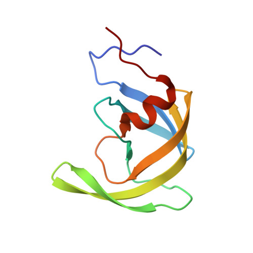
    Mutations in HIV-1 protease (PR) that produce resistance to antiviral PR inhibitors are a major problem in AIDS therapy. The mutation F53L arising from antiretroviral therapy was introduced into the flexible flap region of the wild-type PR to study its effect and potential role in developing drug resistance. Compared to wild-type PR, PR(F53L) showed lower (15%) catalytic efficiency, 20-fold weaker inhibition by the clinical drug indinavir, and reduced dimer stability, while the inhibition constants of two peptide analog inhibitors were slightly lower than those for PR. The crystal structure of PR(F53L) was determined in the unliganded form at 1.35 Angstrom resolution in space group P4(1)2(1)2. The tips of the flaps in PR(F53L) had a wider separation than in unliganded wild-type PR, probably due to the absence of hydrophobic interactions of the side-chains of Phe53 and Ile50'. The changes in interactions between the flaps agreed with the reduced stability of PR(F53L) relative to wild-type PR. The altered flap interactions in the unliganded form of PR(F53L) suggest a distinct mechanism for drug resistance, which has not been observed in other common drug-resistant mutants.


  • Organizational Affiliation

    Department of Biology, Molecular Basis of Disease Program, Georgia State University, Atlanta, GA 30303, USA.


Macromolecules
Find similar proteins by:  (by identity cutoff)  |  3D Structure
Entity ID: 1
MoleculeChains Sequence LengthOrganismDetailsImage
protease99Human immunodeficiency virus 1Mutation(s): 6 
EC: 3.4.23.16
UniProt
Find proteins for Q7SSI0 (Human immunodeficiency virus 1)
Explore Q7SSI0 
Go to UniProtKB:  Q7SSI0
Entity Groups  
Sequence Clusters30% Identity50% Identity70% Identity90% Identity95% Identity100% Identity
UniProt GroupQ7SSI0
Sequence Annotations
Expand
  • Reference Sequence
Experimental Data & Validation

Experimental Data

  • Method: X-RAY DIFFRACTION
  • Resolution: 1.35 Å
  • R-Value Free: 0.234 
  • R-Value Work: 0.155 
  • R-Value Observed: 0.160 
  • Space Group: P 41 21 2
Unit Cell:
Length ( Å )Angle ( ˚ )
a = 60.946α = 90
b = 60.946β = 90
c = 55.551γ = 90
Software Package:
Software NamePurpose
SHELXmodel building
SHELXL-97refinement
MAR345data collection
SCALEPACKdata scaling
CCP4phasing

Structure Validation

View Full Validation Report


Entry History 

Deposition Data

Revision History  (Full details and data files)

  • Version 1.0: 2006-05-09
    Type: Initial release
  • Version 1.1: 2008-05-01
    Changes: Version format compliance
  • Version 1.2: 2011-07-13
    Changes: Derived calculations, Version format compliance
  • Version 1.3: 2017-10-18
    Changes: Refinement description
  • Version 1.4: 2021-10-20
    Changes: Database references
  • Version 1.5: 2024-02-14
    Changes: Data collection