2FZJ

New Insights into DHFR Interactions: Analysis of Pneumocystis carinii and Mouse DHFR Complexes with NADPH and Two Highly Potent Trimethoprim Derivatives


Experimental Data Snapshot

  • Method: X-RAY DIFFRACTION
  • Resolution: 2.00 Å
  • R-Value Free: 0.294 
  • R-Value Work: 0.234 
  • R-Value Observed: 0.234 

wwPDB Validation   3D Report Full Report


Ligand Structure Quality Assessment 


This is version 1.3 of the entry. See complete history


Literature

New insights into DHFR interactions: analysis of Pneumocystis carinii and mouse DHFR complexes with NADPH and two highly potent 5-(omega-carboxy(alkyloxy) trimethoprim derivatives reveals conformational correlations with activity and novel parallel ring stacking interactions.

Cody, V.Pace, J.Chisum, K.Rosowsky, A.

(2006) Proteins 65: 959-969

  • DOI: https://doi.org/10.1002/prot.21131
  • Primary Citation of Related Structures:  
    2FZH, 2FZI, 2FZJ

  • PubMed Abstract: 

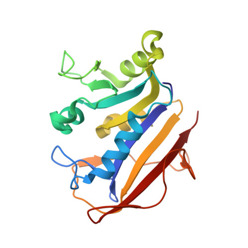
    Structural data are reported for two highly potent antifolates, 2,4-diamino-5-[3',4'-dimethoxy-5'-(5-carboxy-1-pentynyl)]benzylpyrimidine (PY1011), with 5000-fold selectivity for Pneumocystis carinii dihydrofolate reductase (pcDHFR), relative to rat liver DHFR, and 2,4-diamino-5-[2-methoxy-5-(4-carboxybutyloxy)benzyl]pyrimidine (PY957), that has 80-fold selectivity for pcDHFR. Crystal structures are reported for NADPH ternary complexes with PY957 and pcDHFR, refined to 2.2 A resolution; with PY1011 and pcDHFR, refined to 2.0 A resolution; and with PY1011 and mouse DHFR (mDHFR), refined to 2.2 A resolution. These results reveal that the carboxylate of the omega-carboxyalkyloxy side chain of these inhibitors form ionic interactions with the conserved Arg in the substrate binding pocket of DHFR. These data suggest that the enhanced inhibitory activity of PY1011 compared with PY957 is, in part, due to the favorable contacts with Phe69 of pcDHFR by the methylene carbons of the inhibitor side chain that are oriented by the triple bond of the 1-pentynyl side chain. These contacts are not present in the PY957 pcDHFR complex, or in the PY1011 mDHFR complex. In the structure of mDHFR the site of Phe69 in pcDHFR is occupied by Asn64. These data also revealed a preference for an unusual parallel ring stacking interaction between Tyr35 of the active site helix and Phe199 of the C-terminal beta sheet in pcDHFR and by Tyr33 and Phe179 in mDHFR that is independent of bound ligand. A unique His174-His187 parallel ring stacking interaction was also observed only in the structure of pcDHFR. These ring stacking interactions are rarely found in any other protein families and may serve to enhance protein stability.


  • Organizational Affiliation

    Department of Structural Biology, Hauptman-Woodward Medical Research Institute, Buffalo, New York 14203, USA. cody@hwi.buffalo.edu


Macromolecules
Find similar proteins by:  (by identity cutoff)  |  3D Structure
Entity ID: 1
MoleculeChains Sequence LengthOrganismDetailsImage
Dihydrofolate reductase186Mus musculusMutation(s): 0 
Gene Names: Dhfr
EC: 1.5.1.3
UniProt & NIH Common Fund Data Resources
Find proteins for P00375 (Mus musculus)
Explore P00375 
Go to UniProtKB:  P00375
IMPC:  MGI:94890
Entity Groups  
Sequence Clusters30% Identity50% Identity70% Identity90% Identity95% Identity100% Identity
UniProt GroupP00375
Sequence Annotations
Expand
  • Reference Sequence
Small Molecules
Ligands 2 Unique
IDChains Name / Formula / InChI Key2D Diagram3D Interactions
NDP
Query on NDP

Download Ideal Coordinates CCD File 
B [auth A]NADPH DIHYDRO-NICOTINAMIDE-ADENINE-DINUCLEOTIDE PHOSPHATE
C21 H30 N7 O17 P3
ACFIXJIJDZMPPO-NNYOXOHSSA-N
DH3
Query on DH3

Download Ideal Coordinates CCD File 
C [auth A]2,4-DIAMINO-5-[3',4'-DIMETHOXY-5'-(5-CARBOXYL-1-PENTYNYL)]BENZYL PYRIMIDINE
C19 H22 N4 O4
JAGSWKLZBKKKJP-UHFFFAOYSA-N
Binding Affinity Annotations 
IDSourceBinding Affinity
DH3 BindingDB:  2FZJ IC50: 5000 (nM) from 1 assay(s)
Experimental Data & Validation

Experimental Data

  • Method: X-RAY DIFFRACTION
  • Resolution: 2.00 Å
  • R-Value Free: 0.294 
  • R-Value Work: 0.234 
  • R-Value Observed: 0.234 
  • Space Group: P 1 21 1
Unit Cell:
Length ( Å )Angle ( ˚ )
a = 41.806α = 90
b = 61.034β = 118
c = 43.08γ = 90
Software Package:
Software NamePurpose
DENZOdata reduction
SCALEPACKdata scaling
CNSrefinement
CNSphasing

Structure Validation

View Full Validation Report


Ligand Structure Quality Assessment 


Entry History 

Deposition Data

Revision History  (Full details and data files)

  • Version 1.0: 2006-12-26
    Type: Initial release
  • Version 1.1: 2008-05-01
    Changes: Version format compliance
  • Version 1.2: 2011-07-13
    Changes: Version format compliance
  • Version 1.3: 2023-08-30
    Changes: Data collection, Database references, Derived calculations, Refinement description