2FYS

Crystal structure of Erk2 complex with KIM peptide derived from MKP3


Experimental Data Snapshot

  • Method: X-RAY DIFFRACTION
  • Resolution: 2.50 Å
  • R-Value Free: 0.266 
  • R-Value Work: 0.174 

wwPDB Validation   3D Report Full Report


This is version 1.3 of the entry. See complete history


Literature

Structural basis of docking interactions between ERK2 and MAP kinase phosphatase 3

Liu, S.Sun, J.P.Zhou, B.Zhang, Z.Y.

(2006) Proc Natl Acad Sci U S A 103: 5326-5331

  • DOI: https://doi.org/10.1073/pnas.0510506103
  • Primary Citation of Related Structures:  
    2FYS

  • PubMed Abstract: 

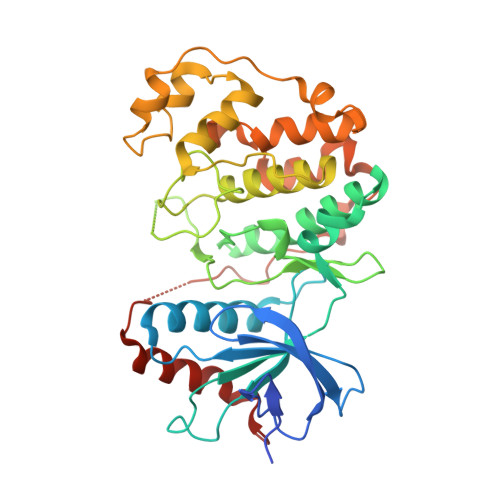
    Mitogen-activated protein (MAP) kinases are central components of signal transduction pathways for cell proliferation, stress responses, and differentiation. Signaling efficiency and specificity are modulated in large part by docking interactions between individual MAP kinase and the kinase interaction motif (KIM), (R/K)(2-3)-X(1-6)-Phi(A)-X-Phi(B), in its cognate kinases, phosphatases, scaffolding proteins, and substrates. We have determined the crystal structure of extracellular signal-regulated protein kinase 2 bound to the KIM peptide from MAP kinase phosphatase 3, an extracellular signal-regulated protein kinase 2-specific phosphatase. The structure reveals that the KIM docking site, situated in a noncatalytic region opposite of the kinase catalytic pocket, is comprised of a highly acidic patch and a hydrophobic groove, which engage the basic and Phi(A)-X-Phi(B) residues, respectively, in the KIM sequence. The specific docking interactions observed in the structure consolidate all known biochemical data. In addition, structural comparison indicates that the KIM docking site is conserved in all MAP kinases. The results establish a structural model for understanding how MAP kinases interact with their regulators and substrates and provide new insights into how MAP kinase docking specificity can be achieved.


  • Organizational Affiliation

    Department of Biochemistry and Molecular Biology, Indiana University School of Medicine, 635 Barnhill Drive, Indianapolis, IN 46202, USA.


Macromolecules
Find similar proteins by:  (by identity cutoff)  |  3D Structure
Entity ID: 1
MoleculeChains Sequence LengthOrganismDetailsImage
Mitogen-activated protein kinase 1A [auth B],
B [auth A]
364Rattus norvegicusMutation(s): 0 
Gene Names: Mapk1Erk2MapkPrkm1
EC: 2.7.1.37
UniProt
Find proteins for P63086 (Rattus norvegicus)
Explore P63086 
Go to UniProtKB:  P63086
Entity Groups  
Sequence Clusters30% Identity50% Identity70% Identity90% Identity95% Identity100% Identity
UniProt GroupP63086
Sequence Annotations
Expand
  • Reference Sequence

Find similar proteins by:  Sequence   |   3D Structure  

Entity ID: 2
MoleculeChains Sequence LengthOrganismDetailsImage
Dual specificity protein phosphatase 6C [auth D],
D [auth C]
17N/AMutation(s): 0 
EC: 3.1.3.48 (PDB Primary Data), 3.1.3.16 (PDB Primary Data)
UniProt
Find proteins for Q64346 (Rattus norvegicus)
Explore Q64346 
Go to UniProtKB:  Q64346
Entity Groups  
Sequence Clusters30% Identity50% Identity70% Identity90% Identity95% Identity100% Identity
UniProt GroupQ64346
Sequence Annotations
Expand
  • Reference Sequence
Experimental Data & Validation

Experimental Data

  • Method: X-RAY DIFFRACTION
  • Resolution: 2.50 Å
  • R-Value Free: 0.266 
  • R-Value Work: 0.174 
  • Space Group: P 1 21 1
Unit Cell:
Length ( Å )Angle ( ˚ )
a = 57.377α = 90
b = 67.478β = 99.55
c = 86.605γ = 90
Software Package:
Software NamePurpose
DENZOdata reduction
SCALEPACKdata scaling
AMoREphasing
CNSrefinement

Structure Validation

View Full Validation Report


Entry History 

Deposition Data

Revision History  (Full details and data files)

  • Version 1.0: 2006-04-11
    Type: Initial release
  • Version 1.1: 2008-05-01
    Changes: Version format compliance
  • Version 1.2: 2011-07-13
    Changes: Version format compliance
  • Version 1.3: 2023-08-30
    Changes: Data collection, Database references, Refinement description