Structure of Botulinum Neurotoxin Type D Light Chain at 1.65 A Resolution: Repercussions for VAMP-2 Substrate Specificity(,).
Arndt, J.W., Chai, Q., Christian, T., Stevens, R.C.(2006) Biochemistry 45: 3255-3262
- PubMed: 16519520
- DOI: https://doi.org/10.1021/bi052518r
- Primary Citation of Related Structures:
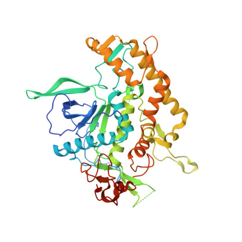
2FPQ - PubMed Abstract:
The seven serotypes (A-G) of botulinum neurotoxins (BoNTs) function through their proteolytic cleavage of one of three proteins (SNAP-25, Syntaxin, and VAMP) that form the SNARE complex required for synaptic vesicle fusion. The different BoNTs have very specific protease recognition requirements, between 15 and 50 amino acids in length depending on the serotype. However, the structural details involved in substrate recognition remain largely unknown. Here is reported the 1.65 A resolution crystal structure of the catalytic domain of BoNT serotype D (BoNT/D-LC), providing insight into the protein-protein binding interaction and final proteolysis of VAMP-2. Structural analysis has identified a hydrophobic pocket potentially involved in substrate recognition of the P1' VAMP residue (Leu 60) and a second remote site for recognition of the V1 SNARE motif that is critical for activity. A structural comparison of BoNT/D-LC with BoNT/F-LC that also recognizes VAMP-2 one residue away from the BoNT/D-LC site provides additional molecular details about the unique serotype specific activities. In particular, BoNT/D prefers a hydrophobic interaction for the V1 motif of VAMP-2, while BoNT/F adopts a more hydrophilic strategy for recognition of the same V1 motif.
Organizational Affiliation:
Department of Molecular Biology, The Scripps Research Institute, 10550 North Torrey Pines Road, La Jolla, California 92037, USA.