2FH5

The Structure of the Mammalian SRP Receptor


Experimental Data Snapshot

  • Method: X-RAY DIFFRACTION
  • Resolution: 2.45 Å
  • R-Value Free: 0.232 
  • R-Value Work: 0.193 
  • R-Value Observed: 0.193 

wwPDB Validation   3D Report Full Report


Ligand Structure Quality Assessment 


This is version 1.2 of the entry. See complete history


Literature

The structure of the mammalian signal recognition particle (SRP) receptor as prototype for the interaction of small GTPases with Longin domains.

Schlenker, O.Hendricks, A.Sinning, I.Wild, K.

(2006) J Biol Chem 281: 8898-8906

  • DOI: https://doi.org/10.1074/jbc.M512415200
  • Primary Citation of Related Structures:  
    2FH5

  • PubMed Abstract: 

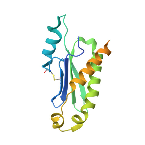
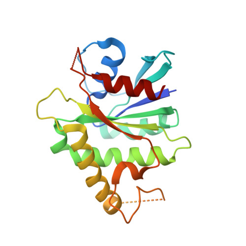
    The eukaryotic signal recognition particle (SRP) and its receptor (SR) play a central role in co-translational targeting of secretory and membrane proteins to the endoplasmic reticulum. The SR is a heterodimeric complex assembled by the two GTPases SRalpha and SRbeta, which is membrane-anchored. Here we present the 2.45-A structure of mammalian SRbeta in its Mg2+ GTP-bound state in complex with the minimal binding domain of SRalpha termed SRX. SRbeta is a member of the Ras-GTPase superfamily closely related to Arf and Sar1, while SRX belongs to the SNARE-like superfamily with a fold also known as longin domain. SRX binds to the P loop and the switch regions of SRbeta-GTP. The binding mode and structural similarity with other GTPase-effector complexes suggests a co-GAP (GTPase-activating protein) function for SRX. Comparison with the homologous yeast structure and other longin domains reveals a conserved adjustable hydrophobic surface within SRX which is of central importance for the SRbeta-GTP:SRX interface. A helix swap in SRX results in the formation of a dimer in the crystal structure. Based on structural conservation we present the SRbeta-GTP:SRX structure as a prototype for conserved interactions in a variety of GTPase regulated targeting events occurring at endomembranes.


  • Organizational Affiliation

    Biochemie-Zentrum der Universität Heidelberg (BZH), Im Neuenheimer Feld 328, D-69120 Heidelberg, Germany.


Macromolecules
Find similar proteins by:  (by identity cutoff)  |  3D Structure
Entity ID: 1
MoleculeChains Sequence LengthOrganismDetailsImage
Signal recognition particle receptor alpha subunit185Homo sapiensMutation(s): 0 
Gene Names: SRPR
UniProt & NIH Common Fund Data Resources
Find proteins for P08240 (Homo sapiens)
Explore P08240 
Go to UniProtKB:  P08240
PHAROS:  P08240
GTEx:  ENSG00000182934 
Entity Groups  
Sequence Clusters30% Identity50% Identity70% Identity90% Identity95% Identity100% Identity
UniProt GroupP08240
Sequence Annotations
Expand
  • Reference Sequence
Find similar proteins by:  (by identity cutoff)  |  3D Structure
Entity ID: 2
MoleculeChains Sequence LengthOrganismDetailsImage
Signal recognition particle receptor beta subunit214Mus musculusMutation(s): 0 
Gene Names: Srprb
UniProt & NIH Common Fund Data Resources
Find proteins for P47758 (Mus musculus)
Explore P47758 
Go to UniProtKB:  P47758
IMPC:  MGI:102964
Entity Groups  
Sequence Clusters30% Identity50% Identity70% Identity90% Identity95% Identity100% Identity
UniProt GroupP47758
Sequence Annotations
Expand
  • Reference Sequence
Small Molecules
Ligands 2 Unique
IDChains Name / Formula / InChI Key2D Diagram3D Interactions
GTP
Query on GTP

Download Ideal Coordinates CCD File 
D [auth B]GUANOSINE-5'-TRIPHOSPHATE
C10 H16 N5 O14 P3
XKMLYUALXHKNFT-UUOKFMHZSA-N
MG
Query on MG

Download Ideal Coordinates CCD File 
C [auth B]MAGNESIUM ION
Mg
JLVVSXFLKOJNIY-UHFFFAOYSA-N
Experimental Data & Validation

Experimental Data

  • Method: X-RAY DIFFRACTION
  • Resolution: 2.45 Å
  • R-Value Free: 0.232 
  • R-Value Work: 0.193 
  • R-Value Observed: 0.193 
  • Space Group: I 2 2 2
Unit Cell:
Length ( Å )Angle ( ˚ )
a = 68.092α = 90
b = 118.348β = 90
c = 120.413γ = 90
Software Package:
Software NamePurpose
CNSrefinement
DENZOdata reduction
SCALEPACKdata scaling
AMoREphasing

Structure Validation

View Full Validation Report


Ligand Structure Quality Assessment 


Entry History 

Deposition Data

Revision History  (Full details and data files)

  • Version 1.0: 2006-01-31
    Type: Initial release
  • Version 1.1: 2008-05-01
    Changes: Version format compliance
  • Version 1.2: 2011-07-13
    Changes: Version format compliance