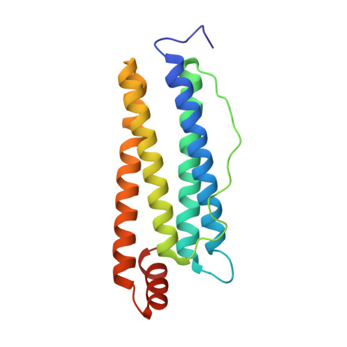
2FG4

Structure of Human Ferritin L Chain


Experimental Data Snapshot

  • Method: X-RAY DIFFRACTION
  • Resolution: 2.10 Å
  • R-Value Free: 0.220 
  • R-Value Work: 0.194 

wwPDB Validation   3D Report Full Report


This is version 1.4 of the entry. See complete history


Literature

Structure of human ferritin L chain.

Wang, Z.Li, C.Ellenburg, M.Soistman, E.Ruble, J.Wright, B.Ho, J.X.Carter, D.C.

(2006) Acta Crystallogr D Biol Crystallogr 62: 800-806

  • DOI: https://doi.org/10.1107/S0907444906018294
  • Primary Citation of Related Structures:  
    2FFX, 2FG4, 2FG8

  • PubMed Abstract: 

    Ferritin is the major iron-storage protein present in all cells. It generally contains 24 subunits, with different ratios of heavy chain (H) to light chain (L), in the shape of a hollow sphere hosting up to 4500 ferric Fe atoms inside. H-rich ferritins catalyse the oxidation of iron(II), while L-rich ferritins promote the nucleation and storage of iron(III). Several X-ray structures have been determined, including those of L-chain ferritins from horse spleen (HoSF), recombinant L-chain ferritins from horse (HoLF), mouse (MoLF) and bullfrog (BfLF) as well as recombinant human H-chain ferritin (HuHF). Here, structures have been determined of two crystal forms of recombinant human L-chain ferritin (HuLF) obtained from native and perdeuterated proteins. The structures show a cluster of acidic residues at the ferrihydrite nucleation site and at the iron channel along the threefold axis. An ordered Cd2+ structure is observed within the iron channel, offering further insight into the route and mechanism of iron transport into the capsid. The loop between helices D and E, which is disordered in many other L-chain structures, is clearly visible in these two structures. The crystals generated from perdeuterated HuLF will be used for neutron diffraction studies.


  • Organizational Affiliation

    New Century Pharmaceuticals Inc., 895 Martin Road, Huntsville, Alabama 35824, USA.


Macromolecules
Find similar proteins by:  (by identity cutoff)  |  3D Structure
Entity ID: 1
MoleculeChains Sequence LengthOrganismDetailsImage
Ferritin light chain174Homo sapiensMutation(s): 0 
Gene Names: FTL
UniProt & NIH Common Fund Data Resources
Find proteins for P02792 (Homo sapiens)
Explore P02792 
Go to UniProtKB:  P02792
PHAROS:  P02792
GTEx:  ENSG00000087086 
Entity Groups  
Sequence Clusters30% Identity50% Identity70% Identity90% Identity95% Identity100% Identity
UniProt GroupP02792
Sequence Annotations
Expand
  • Reference Sequence
Small Molecules
Ligands 1 Unique
IDChains Name / Formula / InChI Key2D Diagram3D Interactions
CD
Query on CD

Download Ideal Coordinates CCD File 
B [auth A]
C [auth A]
D [auth A]
E [auth A]
F [auth A]
B [auth A],
C [auth A],
D [auth A],
E [auth A],
F [auth A],
G [auth A],
H [auth A],
I [auth A],
J [auth A],
K [auth A],
L [auth A],
M [auth A],
N [auth A]
CADMIUM ION
Cd
WLZRMCYVCSSEQC-UHFFFAOYSA-N
Experimental Data & Validation

Experimental Data

  • Method: X-RAY DIFFRACTION
  • Resolution: 2.10 Å
  • R-Value Free: 0.220 
  • R-Value Work: 0.194 
  • Space Group: I 4 3 2
Unit Cell:
Length ( Å )Angle ( ˚ )
a = 152.865α = 90
b = 152.865β = 90
c = 152.865γ = 90
Software Package:
Software NamePurpose
CNXrefinement
DENZOdata reduction
SCALEPACKdata scaling
AMoREphasing

Structure Validation

View Full Validation Report


Entry History 

Deposition Data

Revision History  (Full details and data files)

  • Version 1.0: 2006-07-04
    Type: Initial release
  • Version 1.1: 2008-05-01
    Changes: Version format compliance
  • Version 1.2: 2011-07-13
    Changes: Derived calculations, Version format compliance
  • Version 1.3: 2011-11-16
    Changes: Atomic model
  • Version 1.4: 2023-08-30
    Changes: Data collection, Database references, Derived calculations, Refinement description