2EW6

Structure of Helicobacter Pylori peptide deformylase in complex with inhibitor


Experimental Data Snapshot

  • Method: X-RAY DIFFRACTION
  • Resolution: 2.20 Å
  • R-Value Free: 0.243 
  • R-Value Work: 0.204 
  • R-Value Observed: 0.204 

wwPDB Validation   3D Report Full Report


Ligand Structure Quality Assessment 


This is version 1.4 of the entry. See complete history


Literature

Peptide deformylase is a potential target for anti-Helicobacter pylori drugs: reverse docking, enzymatic assay, and X-ray crystallography validation

Cai, J.Han, C.Hu, T.Zhang, J.Wu, D.Wang, F.Liu, Y.Ding, J.Chen, K.Yue, J.Shen, X.Jiang, H.

(2006) Protein Sci 15: 2071-2081

  • DOI: https://doi.org/10.1110/ps.062238406
  • Primary Citation of Related Structures:  
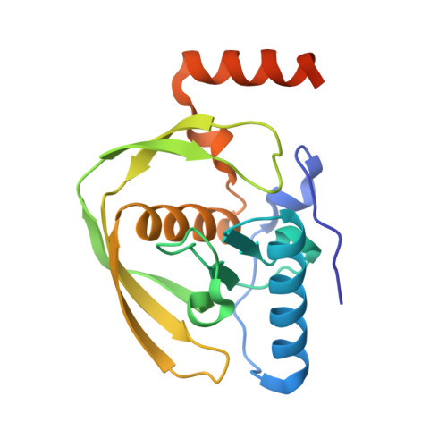
    2EW5, 2EW6, 2EW7

  • PubMed Abstract: 

    Colonization of human stomach by the bacterium Helicobacter pylori is a major causative factor for gastrointestinal illnesses and gastric cancer. However, the discovery of anti-H. pylori agents is a difficult task due to lack of mature protein targets. Therefore, identifying new molecular targets for developing new drugs against H. pylori is obviously necessary. In this study, the in-house potential drug target database (PDTD, http://www.dddc.ac.cn/tarfisdock/) was searched by the reverse docking approach using an active natural product (compound 1) discovered by anti-H. pylori screening as a probe. Homology search revealed that, among the 15 candidates discovered by reverse docking, only diaminopimelate decarboxylase (DC) and peptide deformylase (PDF) have homologous proteins in the genome of H. pylori. Enzymatic assay demonstrated compound 1 and its derivative compound 2 are the potent inhibitors against H. pylori PDF (HpPDF) with IC50 values of 10.8 and 1.25 microM, respectively. X-ray crystal structures of HpPDF and the complexes of HpPDF with 1 and 2 were determined for the first time, indicating that these two inhibitors bind well with HpPDF binding pocket. All these results indicate that HpPDF is a potential target for screening new anti-H. pylori agents. In addition, compounds 1 and 2 were predicted to bind to HpPDF with relatively high selectivity, suggesting they can be used as leads for developing new anti-H. pylori agents. The results demonstrated that our strategy, reverse docking in conjunction with bioassay and structural biology, is effective and can be used as a complementary approach of functional genomics and chemical biology in target identification.


  • Organizational Affiliation

    Drug Discovery and Design Center, State Key Laboratory of Drug Research, Shanghai Institute of Materia Medica Graduate School of Chinese Academy of Sciences, China.


Macromolecules
Find similar proteins by:  (by identity cutoff)  |  3D Structure
Entity ID: 1
MoleculeChains Sequence LengthOrganismDetailsImage
peptide deformylase181Helicobacter pyloriMutation(s): 0 
EC: 3.5.1.88
UniProt
Find proteins for Q672W7 (Helicobacter pylori)
Explore Q672W7 
Go to UniProtKB:  Q672W7
Entity Groups  
Sequence Clusters30% Identity50% Identity70% Identity90% Identity95% Identity100% Identity
UniProt GroupQ672W7
Sequence Annotations
Expand
  • Reference Sequence
Small Molecules
Ligands 2 Unique
IDChains Name / Formula / InChI Key2D Diagram3D Interactions
Y13
Query on Y13

Download Ideal Coordinates CCD File 
C [auth A](2E)-3-(3,4-DIHYDROXYPHENYL)-N-[2-(4-HYDROXYPHENYL)ETHYL]ACRYLAMIDE
C17 H17 N O4
VSHUQLRHTJOKTA-XBXARRHUSA-N
CO
Query on CO

Download Ideal Coordinates CCD File 
B [auth A]COBALT (II) ION
Co
XLJKHNWPARRRJB-UHFFFAOYSA-N
Binding Affinity Annotations 
IDSourceBinding Affinity
Y13 Binding MOAD:  2EW6 IC50: 1.08e+4 (nM) from 1 assay(s)
PDBBind:  2EW6 IC50: 1.08e+4 (nM) from 1 assay(s)
Experimental Data & Validation

Experimental Data

  • Method: X-RAY DIFFRACTION
  • Resolution: 2.20 Å
  • R-Value Free: 0.243 
  • R-Value Work: 0.204 
  • R-Value Observed: 0.204 
  • Space Group: P 21 21 21
Unit Cell:
Length ( Å )Angle ( ˚ )
a = 42.17α = 90
b = 52.937β = 90
c = 91.792γ = 90
Software Package:
Software NamePurpose
CNSrefinement
CCP4data scaling
CNSphasing

Structure Validation

View Full Validation Report


Ligand Structure Quality Assessment 


Entry History 

Deposition Data

  • Released Date: 2006-10-24 
  • Deposition Author(s): Cai, J.

Revision History  (Full details and data files)

  • Version 1.0: 2006-10-24
    Type: Initial release
  • Version 1.1: 2008-05-01
    Changes: Version format compliance
  • Version 1.2: 2011-07-13
    Changes: Version format compliance
  • Version 1.3: 2017-10-18
    Changes: Refinement description
  • Version 1.4: 2023-10-25
    Changes: Data collection, Database references, Derived calculations, Refinement description