2E7E

Bent-binding of cyanide to the heme iron in rat heme oxygenase-1


Experimental Data Snapshot

  • Method: X-RAY DIFFRACTION
  • Resolution: 1.85 Å
  • R-Value Free: 0.218 
  • R-Value Work: 0.194 
  • R-Value Observed: 0.195 

wwPDB Validation   3D Report Full Report


Ligand Structure Quality Assessment 


This is version 1.3 of the entry. See complete history


Literature

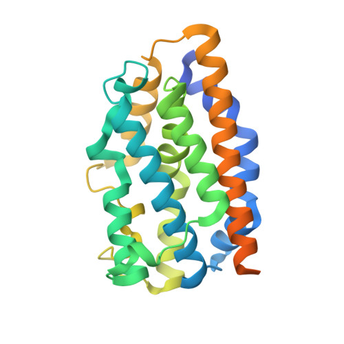
Alternative cyanide-binding modes to the haem iron in haem oxygenase

Sugishima, M.Oda, K.Ogura, T.Sakamoto, H.Noguchi, M.Fukuyama, K.

(2007) Acta Crystallogr Sect F Struct Biol Cryst Commun 63: 471-474

  • DOI: https://doi.org/10.1107/S174430910702475X
  • Primary Citation of Related Structures:  
    2E7E

  • PubMed Abstract: 

    Cyanide is a well known potent inhibitor of haem proteins, including haem oxygenase (HO). Generally, cyanide coordinates to the ferric haem iron with a linear binding geometry; the Fe-C-N angle ranges from 160 to 180 degrees . The Fe-C-N angle observed in the crystal structure of haem-HO bound to cyanide prepared at alkaline pH was 166 degrees . Here, it is reported that cyanide can bind to the haem iron in HO in a bent mode when the ternary complex is prepared at neutral pH; a crystal structure showed that the Fe-C-N angle was bent by 47 degrees . Unlike the ternary complex prepared at alkaline pH, in which the haem group, including the proximal ligand and the distal helix, was displaced upon cyanide binding, the positions of the haem group and the distal helix in the complex prepared at neutral pH were nearly identical to those in haem-HO. Cyanide that was bound to haem-HO with a bent geometry was readily photodissociated, whereas that bound with a linear geometry was not photodissociated. Thus, alternative cyanide-binding modes with linear and bent geometries exist in the crystalline state of haem-HO.


  • Organizational Affiliation

    Department of Medical Biochemistry, Kurume University School of Medicine, Japan.


Macromolecules
Find similar proteins by:  (by identity cutoff)  |  3D Structure
Entity ID: 1
MoleculeChains Sequence LengthOrganismDetailsImage
Heme oxygenase 1267Rattus norvegicusMutation(s): 0 
EC: 1.14.99.3
UniProt
Find proteins for P06762 (Rattus norvegicus)
Explore P06762 
Go to UniProtKB:  P06762
Entity Groups  
Sequence Clusters30% Identity50% Identity70% Identity90% Identity95% Identity100% Identity
UniProt GroupP06762
Sequence Annotations
Expand
  • Reference Sequence
Small Molecules
Ligands 2 Unique
IDChains Name / Formula / InChI Key2D Diagram3D Interactions
HEM
Query on HEM

Download Ideal Coordinates CCD File 
C [auth A]PROTOPORPHYRIN IX CONTAINING FE
C34 H32 Fe N4 O4
KABFMIBPWCXCRK-RGGAHWMASA-L
CYN
Query on CYN

Download Ideal Coordinates CCD File 
B [auth A]CYANIDE ION
C N
XFXPMWWXUTWYJX-UHFFFAOYSA-N
Experimental Data & Validation

Experimental Data

  • Method: X-RAY DIFFRACTION
  • Resolution: 1.85 Å
  • R-Value Free: 0.218 
  • R-Value Work: 0.194 
  • R-Value Observed: 0.195 
  • Space Group: P 32 2 1
Unit Cell:
Length ( Å )Angle ( ˚ )
a = 65.4α = 90
b = 65.4β = 90
c = 121γ = 120
Software Package:
Software NamePurpose
HKL-2000data collection
CNSrefinement
HKL-2000data reduction
HKL-2000data scaling
CNSphasing

Structure Validation

View Full Validation Report


Ligand Structure Quality Assessment 


Entry History 

Deposition Data

Revision History  (Full details and data files)

  • Version 1.0: 2007-06-19
    Type: Initial release
  • Version 1.1: 2008-04-30
    Changes: Version format compliance
  • Version 1.2: 2011-07-13
    Changes: Version format compliance
  • Version 1.3: 2023-10-25
    Changes: Data collection, Database references, Derived calculations, Refinement description