2DHF

CRYSTAL STRUCTURES OF RECOMBINANT HUMAN DIHYDROFOLATE REDUCTASE COMPLEXED WITH FOLATE AND 5-DEAZOFOLATE


Experimental Data Snapshot

  • Method: X-RAY DIFFRACTION
  • Resolution: 2.30 Å
  • R-Value Observed: 0.194 

wwPDB Validation   3D Report Full Report


This is version 1.4 of the entry. See complete history


Literature

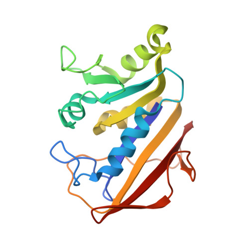
Crystal structures of recombinant human dihydrofolate reductase complexed with folate and 5-deazafolate.

Davies 2nd., J.F.Delcamp, T.J.Prendergast, N.J.Ashford, V.A.Freisheim, J.H.Kraut, J.

(1990) Biochemistry 29: 9467-9479

  • DOI: https://doi.org/10.1021/bi00492a021
  • Primary Citation of Related Structures:  
    1DHF, 2DHF

  • PubMed Abstract: 

    The 2.3-A crystal structure of recombinant human dihydrofolate reductase (EC 1.5.1.3, DHFR) has been solved as a binary complex with folate (a poor substrate at neutral pH) and also as a binary complex with an inhibitor, 5-deazafolate. The inhibitor appears to be protonated at N8 on binding, whereas folate is not. Rotation of the peptide plane joining I7 and V8 from its position in the folate complex permits hydrogen bonding of 5-deazafolate's protonated N8 to the backbone carbonyl of I7, thus contributing to the enzyme's greater affinity for 5-deazafolate than for folate. In this respect it is likely that bound 5-deazafolate furnishes a model for 7,8-dihydrofolate binding and, in addition, resembles the transition state for folate reduction. A hypothetical transition-state model for folate reduction, generated by superposition of the DHFR binary complexes human.5-deazafolate and chicken liver.NADPH, reveals a 1-A overlap of the binding sites for folate's pteridine ring and the dihydronicotinamide ring of NADPH. It is proposed that this binding-site overlap accelerates the reduction of both folate and 7,8-dihydrofolate by simultaneously binding substrate and cofactor with a sub van der Waals separation that is optimal for hydride transfer.


  • Organizational Affiliation

    Department of Chemistry, University of California, San Diego, La Jolla 92093.


Macromolecules
Find similar proteins by:  (by identity cutoff)  |  3D Structure
Entity ID: 1
MoleculeChains Sequence LengthOrganismDetailsImage
DIHYDROFOLATE REDUCTASE
A, B
186Homo sapiensMutation(s): 0 
EC: 1.5.1.3
UniProt & NIH Common Fund Data Resources
Find proteins for P00374 (Homo sapiens)
Explore P00374 
Go to UniProtKB:  P00374
PHAROS:  P00374
GTEx:  ENSG00000228716 
Entity Groups  
Sequence Clusters30% Identity50% Identity70% Identity90% Identity95% Identity100% Identity
UniProt GroupP00374
Sequence Annotations
Expand
  • Reference Sequence
Small Molecules
Ligands 1 Unique
IDChains Name / Formula / InChI Key2D Diagram3D Interactions
DZF
Query on DZF

Download Ideal Coordinates CCD File 
C [auth A],
D [auth B]
5-DEAZAFOLIC ACID
C20 H20 N6 O6
NFARHPAOOHOWAL-AWEZNQCLSA-N
Experimental Data & Validation

Experimental Data

  • Method: X-RAY DIFFRACTION
  • Resolution: 2.30 Å
  • R-Value Observed: 0.194 
  • Space Group: P 1
Unit Cell:
Length ( Å )Angle ( ˚ )
a = 36.1α = 93.9
b = 38.5β = 91.4
c = 76.8γ = 111.5
Software Package:
Software NamePurpose
PROLSQrefinement

Structure Validation

View Full Validation Report


Entry History 

Deposition Data

Revision History  (Full details and data files)

  • Version 1.0: 1990-07-15
    Type: Initial release
  • Version 1.1: 2008-03-24
    Changes: Version format compliance
  • Version 1.2: 2011-07-13
    Changes: Version format compliance
  • Version 1.3: 2017-11-29
    Changes: Derived calculations, Other, Structure summary
  • Version 1.4: 2024-02-14
    Changes: Data collection, Database references, Derived calculations