Identification of the substrate interaction region of the chitin-binding domain of Streptomyces griseus chitinase C
Akagi, K., Watanabe, J., Hara, M., Kezuka, Y., Chikaishi, E., Yamaguchi, T., Akutsu, H., Nonaka, T., Watanabe, T., Ikegami, T.(2006) J Biochem 139: 483-493
- PubMed: 16567413
- DOI: https://doi.org/10.1093/jb/mvj062
- Primary Citation of Related Structures:
2D49 - PubMed Abstract:
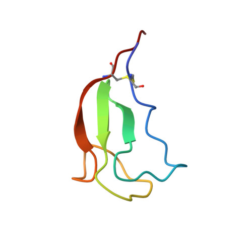
Chitinase C from Streptomyces griseus HUT6037 was discovered as the first bacterial chitinase in family 19 other than chitinases found in higher plants. Chitinase C comprises two domains: a chitin-binding domain (ChBD(ChiC)) for attachment to chitin and a chitin-catalytic domain for digesting chitin. The structure of ChBD(ChiC) was determined by means of 13C-, 15N-, and 1H-resonance nuclear magnetic resonance (NMR) spectroscopy. The conformation of its backbone comprised two beta-sheets composed of two and three antiparallel beta-strands, respectively, this being very similar to the backbone conformations of the cellulose-binding domain of endoglucanase Z from Erwinia chrysanthemi (CBD(EGZ)) and the chitin-binding domain of chitinase A1 from Bacillus circulans WL-12 (ChBD(ChiA1)). The interaction between ChBD(ChiC) and hexa-N-acetyl-chitohexaose was monitored through chemical shift perturbations, which showed that ChBD(ChiC) interacted with the substrate through two aromatic rings exposed to the solvent as CBD(EGZ) interacts with cellulose through three characteristic aromatic rings. Comparison of the conformations of ChBD(ChiA1), ChBD(ChiC), and other typical chitin- and cellulose-binding domains, which have three solvent-exposed aromatic residues responsible for binding to polysaccharides, has suggested that they have adopted versatile binding site conformations depending on the substrates, with almost the same backbone conformations being retained.
Organizational Affiliation:
Laboratory of Structural Proteomics, Institute for Protein Research, Osaka University, 3-2 Yamadaoka, Suita, Osaka 565-0871.