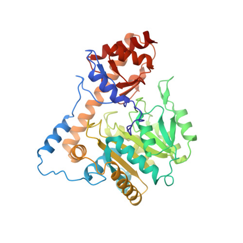
Crystal structure of the closed form of chicken cytosolic aspartate aminotransferase at 1.9 A resolution.
Malashkevich, V.N., Strokopytov, B.V., Borisov, V.V., Dauter, Z., Wilson, K.S., Torchinsky, Y.M.(1995) J Mol Biol 247: 111-124
- PubMed: 7897655
- DOI: https://doi.org/10.1006/jmbi.1994.0126
- Primary Citation of Related Structures:
2CST - PubMed Abstract:
The crystal structure of chicken cytosolic aspartate aminotransferase (cAATase; EC 2.6.1.1) has been solved and refined at 1.9 A resolution. Orthorhombic crystals, space group P2(1)2(1)2(1), a = 56.4 A, b = 126.0 A and c = 142.3 A, were grown from polyethylene glycol solutions in the presence of maleate, a dicarboxylic inhibitor that forms a Michaelis-like complex. The pyridoxal form of the enzyme was used for crystallization. Diffraction data were collected using synchrotron radiation. The structure of the new orthorhombic crystal form was solved by molecular replacement using the partially refined 2.8 A resolution structure of the high-salt crystal form as a search model. The final value of the crystallographic R-factor after rigid body and restrained least-squares refinement is 0.175 with very good model geometry. The two 2-fold-related subunits of cAATase have distinct environments in the crystal lattice. Domain movement is strictly hindered by the lattice contacts in one subunit, while the second one possesses conformational freedom. Despite their different environments, both subunits were found in the closed conformation with one maleate molecule tightly bound in each active site. The present study allows a detailed comparison of the highly refined structures of the aspartate aminotransferase isozymes, and thus provide better insight into the role of conserved and variable residues in substrate recognition and catalysis.
Organizational Affiliation:
Institute of Molecular Biology, Academy of Sciences of the Russia, Moscow.