Structural and Binding Studies of the Three-Metal Center in Two Mycobacterial Ppm Ser/Thr Protein Phosphatases.
Wehenkel, A., Bellinzoni, M., Schaeffer, F., Villarino, A., Alzari, P.M.(2007) J Mol Biol 374: 890
- PubMed: 17961594
- DOI: https://doi.org/10.1016/j.jmb.2007.09.076
- Primary Citation of Related Structures:
2CM1, 2V06 - PubMed Abstract:
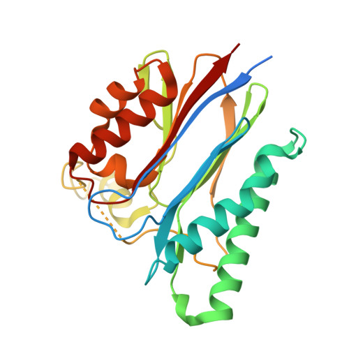
Phospho-Ser/Thr protein phosphatases (PPs) are dinuclear metalloenzymes classed into two large families, PPP and PPM, on the basis of sequence similarity and metal ion dependence. The archetype of the PPM family is the alpha isoform of human PP2C (PP2Calpha), which folds into an alpha/beta domain similar to those of PPP enzymes. The recent structural studies of three bacterial PPM phosphatases, Mycobacterium tuberculosis MtPstP, Mycobacterium smegmatis MspP, and Streptococcus agalactiae STP, confirmed the conservation of the overall fold and dinuclear metal center in the family, but surprisingly revealed the presence of a third conserved metal-binding site in the active site. To gain insight into the roles of the three-metal center in bacterial enzymes, we report structural and metal-binding studies of MtPstP and MspP. The structure of MtPstP in a new trigonal crystal form revealed a fully active enzyme with the canonical dinuclear metal center but without the third metal ion bound to the catalytic site. The absence of metal correlates with a partially unstructured flap segment, indicating that the third manganese ion contributes to reposition the flap, but is dispensable for catalysis. Studies of metal binding to MspP using isothermal titration calorimetry revealed that the three Mn(2+)-binding sites display distinct affinities, with dissociation constants in the nano- and micromolar range for the two catalytic metal ions and a significantly lower affinity for the third metal-binding site. In agreement, the structure of inactive MspP at acidic pH was determined at atomic resolution and shown to lack the third metal ion in the active site. Structural comparisons of all bacterial phosphatases revealed positional variations in the third metal-binding site that are correlated with the presence of bound substrate and the conformation of the flap segment, supporting a role of this metal ion in assisting enzyme-substrate interactions.
Organizational Affiliation:
Unité de Biochimie Structurale and CNRS-URA 2185, Institut Pasteur, 25 rue du Docteur Roux, 75724 Paris Cedex 15, France.