Structure of Cytochrome C(6A), a Novel Dithio-Cytochrome of Arabidopsis Thaliana, and its Reactivity with Plastocyanin: Implications for Function.
Marcaida, M.J., Schlarb-Ridley, B.G., Worrall, J.A.R., Wastl, J., Evans, T.J., Bendall, D.S., Luisi, B.F., Howe, C.J.(2006) J Mol Biol 360: 968
- PubMed: 16815443
- DOI: https://doi.org/10.1016/j.jmb.2006.05.065
- Primary Citation of Related Structures:
2CE0, 2CE1 - PubMed Abstract:
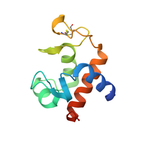
Cytochrome c6A is a unique dithio-cytochrome present in land plants and some green algae. Its sequence and occurrence in the thylakoid lumen suggest that it is derived from cytochrome c6, which functions in photosynthetic electron transfer between the cytochrome b6f complex and photosystem I. Its known properties, however, and a strong indication that the disulfide group is not purely structural, indicate that it has a different, unidentified function. To help in the elucidation of this function the crystal structure of cytochrome c6A from Arabidopsis thaliana has been determined in the two redox states of the heme group, at resolutions of 1.2 A (ferric) and 1.4 A (ferrous). These two structures were virtually identical, leading to the functionally important conclusion that the heme and disulfide groups do not communicate by conformational change. They also show, however, that electron transfer between the reduced disulfide and the heme is feasible. We therefore suggest that the role of cytochrome c6A is to use its disulfide group to oxidize dithiol/disulfide groups of other proteins of the thylakoid lumen, followed by internal electron transfer from the dithiol to the heme, and re-oxidation of the heme by another thylakoid oxidant. Consistent with this model, we found a rapid electron transfer between ferro-cytochrome c6A and plastocyanin, with a second-order rate constant, k2=1.2 x 10(7) M(-1) s(-1).
Organizational Affiliation:
Department of Biochemistry, University of Cambridge, Tennis Court Road, Cambridge CB2 1QW, UK.