2C9C

Structural basis of the nucleotide driven conformational changes in the AAA domain of transcription activator PspF


Experimental Data Snapshot

  • Method: X-RAY DIFFRACTION
  • Resolution: 2.10 Å
  • R-Value Free: 0.198 
  • R-Value Work: 0.160 
  • R-Value Observed: 0.162 

wwPDB Validation   3D Report Full Report


Ligand Structure Quality Assessment 


This is version 1.3 of the entry. See complete history


Literature

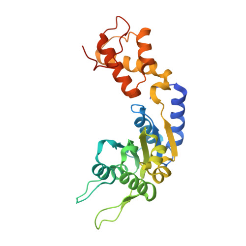
Structural Basis of the Nucleotide Driven Conformational Changes in the Aaa(+) Domain of Transcription Activator Pspf.

Rappas, M.Schumacher, J.Niwa, H.Buck, M.Zhang, X.

(2006) J Mol Biol 357: 481

  • DOI: https://doi.org/10.1016/j.jmb.2005.12.052
  • Primary Citation of Related Structures:  
    2C96, 2C98, 2C99, 2C9C

  • PubMed Abstract: 

    Bacterial enhancer-binding proteins (EBP) activate transcription by hydrolyzing ATP to restructure the sigma(54)-RNA polymerase-promoter complex. We compare six high resolution structures (<2.1 A) of the AAA(+) domain of EBP phage shock protein F (PspF) including apo, AMPPNP, Mg(2+)-ATP, and ADP forms. These structures permit a description of the atomic details underpinning the origins of the conformational changes occurring during ATP hydrolysis. Conserved regions of PspF's AAA(+) domain respond distinctively to nucleotide binding and hydrolysis, suggesting functional roles during the hydrolysis cycle, which completely agree with those derived from activities of PspF mutated at these positions. We propose a putative atomic switch that is responsible for coupling structural changes in the nucleotide-binding site to the repositioning of the sigma(54)-interacting loops. Striking similarities in nucleotide-specific conformational changes and atomic switch exist between PspF and the large T antigen helicase, suggesting conservation in the origin of those events amongst AAA(+) proteins.


  • Organizational Affiliation

    Division of Molecular Biosciences, Imperial College London, London SW7 2AZ, UK.


Macromolecules
Find similar proteins by:  (by identity cutoff)  |  3D Structure
Entity ID: 1
MoleculeChains Sequence LengthOrganismDetailsImage
PSP OPERON TRANSCRIPTIONAL ACTIVATOR265Escherichia coli K-12Mutation(s): 1 
UniProt
Find proteins for P37344 (Escherichia coli (strain K12))
Explore P37344 
Go to UniProtKB:  P37344
Entity Groups  
Sequence Clusters30% Identity50% Identity70% Identity90% Identity95% Identity100% Identity
UniProt GroupP37344
Sequence Annotations
Expand
  • Reference Sequence
Small Molecules
Ligands 2 Unique
IDChains Name / Formula / InChI Key2D Diagram3D Interactions
ATP
Query on ATP

Download Ideal Coordinates CCD File 
C [auth A]ADENOSINE-5'-TRIPHOSPHATE
C10 H16 N5 O13 P3
ZKHQWZAMYRWXGA-KQYNXXCUSA-N
MG
Query on MG

Download Ideal Coordinates CCD File 
B [auth A]MAGNESIUM ION
Mg
JLVVSXFLKOJNIY-UHFFFAOYSA-N
Experimental Data & Validation

Experimental Data

  • Method: X-RAY DIFFRACTION
  • Resolution: 2.10 Å
  • R-Value Free: 0.198 
  • R-Value Work: 0.160 
  • R-Value Observed: 0.162 
  • Space Group: P 65
Unit Cell:
Length ( Å )Angle ( ˚ )
a = 112.243α = 90
b = 112.243β = 90
c = 39.217γ = 120
Software Package:
Software NamePurpose
REFMACrefinement
DENZOdata reduction
SCALEPACKdata scaling
PHASERphasing

Structure Validation

View Full Validation Report


Ligand Structure Quality Assessment 


Entry History 

Deposition Data

Revision History  (Full details and data files)

  • Version 1.0: 2006-02-01
    Type: Initial release
  • Version 1.1: 2011-07-13
    Changes: Advisory, Refinement description, Version format compliance
  • Version 1.2: 2013-03-27
    Changes: Other
  • Version 1.3: 2023-12-13
    Changes: Data collection, Database references, Derived calculations, Other, Refinement description